69,99 €
Mehr erfahren.
- Herausgeber: Thieme
- Kategorie: Fachliteratur
- Sprache: Englisch
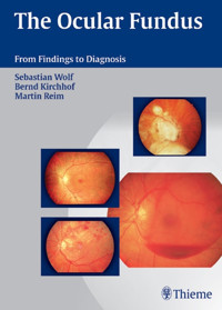
Learn how to "read" the optic fundus: What tests are indicated? How do I interpret the findings? What is the next step? This book guides you quickly and confidently from finding to diagnosis. Practice-oriented Organized by presentation Systematic listing of diagnoses for each presentation Sidebars with a brief summary of the signs and symptoms for each diagnosis Quick reference and study guide in one Comprehensive Describes various examination method Covers even rare findings Differential diagnosis Figures to illustrate each diagnosis Notes on appropriate treatment Confidence Learn to take prompt, goal-directed action. Apply various diagnostic options appropriately and economically. Gain confidence in dealing with equivocal findings.
Das E-Book können Sie in Legimi-Apps oder einer beliebigen App lesen, die das folgende Format unterstützen:
Seitenzahl: 430
Veröffentlichungsjahr: 2005
Ähnliche


Library of Congress Cataloging-in-Publication Data
Wolf, Sebastian.
[Diagnosen am Augenhintergrund. English]
The ocular fundus : from findings to diagnosis / Sebastian Wolf,
Bernd Kirchhof, Martin Reim.
p. ; cm.
Includes bibliographical references and index.
ISBN 3-13-139371-8 (GTV : alk. paper) – ISBN 1-58890-338-9
(TNY : alk. paper)
Eye–Diseases–Diagnosis.
Therapeutics, Ophthalmological. [DNLM: 1. Eye Diseases–diagnosis.
Diagnostic Techniques, Ophthalmological.
Fundus Oculi. WW 141 W855d 2006]
Kirchhof, Bernd.
Reim, Martin.
Title.
RE46.W83515 2006
2005022854
617.7'154–dc22
This book is an authorized and revised translation of the German edition published and copyrighted 2004 by Georg Thieme Verlag, Stuttgart, Germany. Title of the German edition: Diagnosen am Augenhintergrund – Vom Befund zur Diagnose
Translator: John Grossman, M.A., Berlin, Germany Illustrator: E.W. Hanns, Schriesheim
© 2006 Georg Thieme Verlag Rüdigerstraße 14 D-70469 Stuttgart, Germany http://www.thieme.de
Thieme New York, 333 Seventh Avenue, New York, NY 10001 USA http://www.thieme.com
Typesetting by OADF, Altdorf. www.oadf.de Printed in Germany by Appl, Wemding
ISBN 3-13-139371-8 (GTV)
ISBN 1-58890-338-9 (TNY)
123456
Important note: Medicine is an ever-changing science undergoing continual development. Research and clinical experience are continually expanding our knowledge, in particular our knowledge of proper treatment and drug therapy. Insofar as this book mentions any dosage or application, readers may rest assured that the authors, editors, and publishers have made every effort to ensure that such references are in accordance with the state of knowledge at the time of production of the book.
Nevertheless, this does not involve, imply, or express any guarantee or responsibility on the part of the publishers in respect to any dosage instructions and forms of applications stated in the book. Every user is requested to examine carefully the manufacturers' leaflets accompanying each drug and to check, if necessary in consultation with a physician or specialist, whether the dosage schedules mentioned therein or the contraindications stated by the manufacturers differ from the statements made in the present book. Such examination is particularly important with drugs that are either rarely used or have been newly released on the market. Every dosage schedule or every form of application used is entirely at the user's own risk and responsibility. The authors and publishers request every user to report to the publishers any discrepancies or inaccuracies noticed. If errors in this work are found after publication, errata will be posted at www.thieme.com on the product description page.
Some of the product names, patents, and registered designs referred to in this book are in fact registered trademarks or proprietary names even though specific reference to this fact is not always made in the text. Therefore, the appearance of a name without designation as proprietary is not to be construed as a representation by the publisher that it is in the public domain.
This book, including all parts thereof, is legally protected by copyright. Any use, exploitation, or commercialization outside the narrow limits set by copyright legislation, without the publisher's consent, is illegal and liable to prosecution. This applies in particular to photostat reproduction, copying, mimeographing, preparation of microfilms, and electronic data processing and storage.
Preface
Thorough clinical examinations and meticulous documentation of findings continue to provide the basis for diagnosis and treatment in ophthalmologic practice. Fundus photography helps to objectively document findings. It also allows retrospective analysis and discussion of clinical syndromes at a later date. This book represents a compilation of decades of experience in the examination and treatment of ophthalmic disorders in clinical practice. We were motivated to undertake this project by our observation that uncertainty on the part of the examiner in evaluating fundus findings coupled with a tendency to fall back on relatively few diagnoses is a common and recurrent problem. This book is intended to provide a concise yet broad-based overview, ranging from the definition of simple fundus changes to detailed presentations of clinical syndromes.
In contrast to other systematically structured textbooks, we have chosen to follow a somewhat unconventional concept and have organized the material according to symptoms. This approach is intended to ease the transition from findings to diagnosis. It allows us to discuss considerations that are important for a differential diagnosis.
The development of new examination methods has gradually extended the ophthalmologist's diagnostic repertoire and has opened up realms that are otherwise inaccessible by ophthalmoscopic visualization. With its advent in 1972, fluorescein angiography expanded the range of diagnostic modalities for disorders of the retina and choroid. This once novel technique has increased the interest of many ophthalmologists in fundus findings, which are often difficult to interpret. At the same time, the advent of computer technology has simplified specific electrophysiologic examinations.
Video fluorescein angiography was developed by Norbert Körber and Sebastian Wolf at the Eye Clinic of RWTH Aachen Medical School in Germany in the late 1970s and early 1980s. Sebastian Wolf's programming of one of the first scanning laser ophthalmoscopes for clinical use introduced a new dimension in clinical fundus angiography. Around 1990, Wolf used scanning laser ophthalmoscopy to demonstrate the clinical value of choroid angiography with indocyanine green, leading to major advances in the diagnosis of age-related macular degeneration. Multifocal electroretinography and optic computed tomography (OCT) made their appearance at nearly the same time. Finally, Doppler ultrasound examinations of the orbital, choroidal, and retinal vessels supplemented video angiographic measurements of blood flow in the retina and optic disc.
The Eye Clinic of RWTH Aachen has enjoyed a very favorable constellation for ophthalmic examinations for over two decades. In addition to having a staff of highly dedicated and interested ophthalmologists, fully operational units for angiographic, electrophysiologic, and ultrasound examinations have been available continuously. With its capabilities for fundus photography and digital image documentation, the Eye Clinic's photographic department has been an invaluable resource. Monika Bindewald, working in the 1970s, and Willebald Te Poel, active since 1980, have produced valuable image material for this book.
The ongoing collaboration with my wife, Hildegard Reim, MD, and Professor Sebastian Wolf, MD, DSc, has been indispensable and immensely constructive. They promptly processed findings from their daily clinical practice and presented many of them at the Eye Clinic's daily staff conferences. Notable cases were also presented for discussion at the semiannual meeting of the international FAN Club (Fluorescein Angiography Netherlands) and published in medical literature.
Another fortunate circumstance for the production of this book was the fact that Professor Bernd Kirchhof, MD, became director of the Eye Clinic in Aachen in 1996, bringing with him the experience of the large university eye clinic in Cologne. His beneficial activities at the Eye Clinic in Aachen contributed immensely to the success of this work.
As senior author, it is my duty to thank all those who have contributed to the compilation of this material for the manuscript and the extensive imaging documentation. My recognition applies not only to those mentioned above, but also to the many assistants and attending physicians of the Eye Hospital in Aachen who have contributed to the hospital's collection of training material.
I would like to express my heartfelt thanks to my coauthors. We have fond memories of the author conferences that gave rise to the concept and content of this book. The two active clinicians have earned very special thanks for their tireless cooperation.
We are very grateful to our publisher, Georg Thieme Verlag, for the arduous and meticulous work involved in producing the German edition of this book. Without the expertise and efforts of Dr Michael Wachinger, Antje-Karen Richter, Mona Chatterjee, Rolf Zeller, and other unnamed Thieme employees, the manuscript would never have taken on its present form, and the image documentation would have continued to rest in the deep drawers of the photo archive. We would also like to thank Dr Clifford Bergman and Gabriele Kuhn of editorial, and Stefanie Langner of production, for their competent management of the English edition, as well as the translator himself, John Grossman, for his accurate and lucid translation of the original.
We now wish the book many curious readers. As with any comprehensive printed work, and especially in the first edition, errors on the part of the authors or publisher may occur. We are grateful to readers who notify us of any such oversights.
Sebastian Wolf, Bern
Bernd Kirchhof, Cologne
Martin Reim, Aachen
Table of Contents
1 Introduction
2 Examination Methods
History
Functional Tests
Visual Acuity
Visual Field
Color Vision
Contrast Sensitivity
Ophthalmoscopy
Direct Ophthalmoscopy
Indirect Ophthalmoscopy
High Refractive Power Lenses in Conjunction with the Slit Lamp
Contact Optics with Deflecting Mirrors
Special Considerations when Examining Infants and Newborns
Objective Imaging Studies and Their Evaluation
Fundus Photography
Scanning Laser Ophthalmoscope
Fluorescence Angiography
Electrophysiological Studies
Electro-oculography (EOG)
Electroretinography (ERG)
Visual Evoked Cortical Potentials (VECP)
Ultrasound Studies
A-Mode Ultrasonography
B-Mode Ultrasonography
Doppler Ultrasonography
Color Doppler Imaging
Ultrasound Biomicroscopy
Biopsy
Anterior Chamber
Vitreous Body
Tumors
3 Appearance of Retinal and Choroidal Disorders
Retinopathy with Focal or Mottled Lesions
White Mottled Deposits
Sharply Demarcated, Bright, and Pigmented Areas
Blurred, Fluffy, White Focal Lesions
Prominence of the Macula
Secondary Macular Edema
Macular Edema with Retinal Vascular Disease
Macular Edema from Choroidal Neovascularization
Choroidal Neovascularization of Other Causes
Proliferation, Scarring, and Holes in the Macula
Epiretinal Membranes (Macular Pucker)
Idiopathic Macular Hole
Choroidal Folds
Depigmented and Pigmented Focal Lesions in the Macula
Solar Retinopathy
Injuries from Laser Beams and Autogenous Welding
Pigmented Macular Scars Secondary to Contusion
Maculopathy in Myopia
Pattern Dystrophies
Central Areolar Macular Atrophy
Bull’s Eye Dystrophy
Macular Dystrophies with Mottled Lesions
Vitelliform Macular Dystrophy (Best Disease)
Sorsby Macular Dystrophy
Stargardt Disease
Large Areas of Yellowish-White Exudative Retinopathy
Necrotizing Retinitis
Serpiginous Choroiditis
Vogt–Koyanagi–Harada Syndrome
Berlin Edema in the Fundus
Ischemic Edema in Central Retinal Artery Occlusion
Scattered Pigment Changes with Large Areas of Retinal and Choroidal Dystrophy
Atrophic and Pigmented Areas
Fields with Coarse Pigmentation and Depigmentation
Large Areas of Progressive Retinal Degeneration in Both Eyes—Cancer-Associated Retinopathy (CAR)
Significance of Rare Types of Retinal Degeneration and Dystrophy
Fundi with Typical Pigmentations and Chorioretinal Atrophy—Tapetoretinal Degenerations
Albinism
Pediatric Retinal Dystrophies with Maculopathies
Peripheral Retinal and Choroidal Lesions
Pigmentation, Bright White–Yellow Spots and Cobblestone Degeneration
Lattice Degeneration and Vitreous Adhesion
Retinal Holes and Tears
Pigmented Flecks and Fields at the Level of the Fundus
Vitreoretinal Degeneration
Retinal Detachments
Broad Gray Bubblelike Bulging of the Retina
Glassy, Transparent Retinal Detachments
Choroidal Detachment
Retinal Detachment due to Solid Protrusions
Retinal Tumors
White Tumors—Retinoblastoma
“Mulberry” Tumors: Tuberous Sclerosis
Vascular Tumors: Hemangiomas
Brown Tumor in Coats Disease
4 Appearance of Vascular Disorders
Ophthalmoscopic Structure of Fundus Vessels
Variants in the Course of the Retinal Vessels
Cilioretinal Artery
Atypical Branching of the Retinal Arterioles
Retinal Vessels in Macular Aplasia
Abnormal Vessels in the Retina
Telangiectasia
Multiple Leber Miliary Aneurysms
Solitary Retinal Macroaneurysms
Angiomatosis Retinae (Von Hippel–Lindau Disease)
Rarefied and Elongated Vessels
Wide and Irregular Reflexes on Retinal Arterioles in Arteriosclerosis
Choroidal Sclerosis with Bright Vascular Bands and Thin Columns of Blood
Thin Vessels, Minimal Microvasculature, and Pale Fundus in Ischemic Ophthalmopathy
Thin Vessels, Pale Fundus, and Occasional Retinal Bleeding in Anemia
Narrowed Arterioles and Congested Veins
Pathophysiology of the Retinal Vessels in Arterial Hypertension
Changes in the Retinal Vessels in Arterial Hypertension
Distinguishing Arteriosclerosis from Hypertension
Hypertensive Fundus
Bleeding, Cotton-Wool Spots, Hard Exudates, and Retinal Edema
Hypertensive Retinopathy
Classification of Fundus Changes in Arterial Hypertension
Stenoses of the Arterioles, Cotton-Wool Spots, and Retinal Edema in Retinopathy in Pregnancy
Purtscher Retinopathy (Traumatic Retinal Angiopathy)
Capillary Aneurysms, Hard Exudates, Bleeding, and Neovascularization
Nonproliferative Diabetic Retinopathy (NPDR)
Proliferative Diabetic Retinopathy (PDR)
Macular Edema in Diabetic Maculopathy
Disorders Involving Primarily Retinal Bleeding
Bleeding in the Region Drained by the Retinal Veins
Cockade Hemorrhagic Lesions in Chronic Myelosis
Bleeding between the Venules and Arterioles in Waldenström Macroglobulinemia
Large Areas of Bleeding in Multiple Myelomas (Plasmacytoma)
Engorged Blood Vessels in Polycythemia Vera
Idiopathic Macular Bleeding—Macular Bleeding Following the Valsalva Maneuver
Characteristic Fundus Findings in Hemoglobin Disorders
Peripheral Neovascularization in the Retina
Retinopathy of Prematurity (ROP)
Peripheral Vascular Proliferation in Eales Disease
Perivascular Infiltrates, Vascular Obliteration, and Retinal Bleeding in Inflammatory Vascular Disorders
Peripheral Vascular Obliteration in Eales Disease
Perivascular Infiltrates in Retinal Periphlebitis
Inflammatory Infiltration in Specific Types of Retinal Vasculitis
5 Phenomenology of Disease of Vitreous Body
Symptoms with Vitreous Opacities and Specific Examinations
Leukocoria
Blood Vessels in the Vitreous Body
Persistent Hyaloid Artery arising from the Optic Disc
Active Neovascularization in the Vitreous Body in Proliferative Retinopathy
Small Opacities in the Vitreous Body
Floaters—Moving Spots against a Bright Background
Distressing Subjective Perceptions in Posterior Vitreous Detachment
Multiple White Dots like Snow Flakes Synchysis Scintillans—Cholesterol Hyalosis
Multiple White Globules—Asteroid Hyalosis— Scintillatio Nivea
Diffuse Opacities in the Vitreous Body
Opacities from Chronic Cellular and Fibrinous Infiltration of the Vitreous Body
Opacities from Acute Cellular and Fibrinous Infiltration of the Vitreous Body
Large Opacities in the Vitreous Body
Vitreous Hemorrhage
6 Appearance of Optic Nerve Disorders
Blurred Appearance, Hyperemia and Protrusion—Optic Disc Edema
Bilateral Blurring of the Optic Disc with Hyperemia and Prominence
Unilateral Blurring of the Optic Disc with Hyperemia, Prominence, and Impaired Vision
Pale, Often White, Sharply Demarcated Optic Disc—Optic Nerve Atrophy
Diagnosis and Pathogenesis of Optic Nerve Atrophy
Partial or Localized Pallor of the Optic Disc
Completely White Optic Disc— Optic Nerve Atrophy
Hereditary Optic Nerve Atrophy
Excavations of the Optic Nerve
Physiologic Cupping of the Optic Disc
Cupping of the Optic Disc in Myopia
Cupping and Atrophy of the Optic Nerve in Glaucoma
Excavation Filled with Grayish-White Tissue— Optic Pit
Anomalous Tissue on and adjacent to the Optic Disc
Scleral Crescent—Pigment Crescent— Myopic Crescent
Persistent Embryonic Tissue on the Optic Disc
Optic Disc Coloboma—Morning Glory Disc
Optic Disc Melanocytoma
Optic Disc Hemangioma
Optic Disc Osteoma
7 Disorders without Conspicuous Changes of the Fundus
Floaters
Unilateral Visual Impairment in Children—Amblyopia
Acute Visual Impairment with Normal Optic Disc
Retrobulbar Optic Neuritis
Visual Impairment with Normal Optic Disc
Example of Compression Neuropathy of the Optic Nerve—Severe Endocrine Orbitopathy (Graves Disease)
Color Vision Defects
Congenital Hereditary Color Vision Defects
Achromatopsia—Stationary Cone Dysfunction
Incipient Tapetoretinal Degeneration
Incipient Stargardt Disease
Incipient Retinitis Pigmentosa
Congenital Stationary Night Blindness (CSNB)
Night Blindness with Hereditary Deficiency of the Retinol-Binding Protein
Simulation
Visual Agnosia
List of Abbreviations
Literature
Systematic Outline of Fundus Diseases
Index
1 Introduction
The fundus is unanimously understood to include retina, retinal pigment epithelium, and choroid as viewed with conventional ophthalmoscopes. These layers of tissue extend far into the periphery of the fundus, enveloping all but the anterior base of the vitreous body.
Fig. 1.1 Cross-section of the eye.
This book discusses diseases occurring posterior to the diaphragm formed by the iris and lens. This space cannot be seen by the focal illumination of the slit lamp microscope alone. The fundus can only be visualized by the aid of additional optics. This was the exciting new dimension that Hermann von Helmholtz, Albrecht von Graefe, and their students introduced with the invention of the ophthalmoscope in 1851. The fundus image was upright and highly magnified. Disadvantages of direct ophthalmoscopy were the limited field of view and the monocular viewing technique. Ruete's work introduced indirect ophthalmoscopy in 1852, which greatly improved the possibilities for examination.
For a long time, ophthalmologists had to make do with the simple examination technique and relatively weak light sources. It was only in the 1950s that high intensity light bulbs with tiny, nearly punctate incandescent elements became available. In Germany, a convenient, monocular indirect ophthalmoscope was developed by G. Meyer-Schwickerath and K. Bohnen that provided an overview. Fison in England and Schepens in Boston developed binocular indirect ophthalmoscopes, which offered a three-dimensional view of the fundus. These instruments were adapted to a headset, enabling the examiner to have both hands free for surgical procedures. A smaller version of the indirect binocular ophthalmoscope designed by Foerster in Essen became popular in Germany. Mounted on eyeglass frames, the ophthalmoscope is very light and is powered by a small accumulator that the examiner can carry in the pocket of a coat.
In 1948, shortly after the end of the Second World War, Hans Goldmann in Bern, Switzerland, invented the three-mirror contact lens, which allows binocular biomicroscopy of the central and peripheral fundus at high resolution. At the same time, Karl Hruby in Vienna, Austria, introduced the –55-diopter concave lens that bears his name. Mounted on the slit lamp, this optical system allowed noncontact imaging of the fundus. The panfundoscope developed by Schlegel in Homburg, Germany employed a large converging lens (90 diopters) and was applied to the cornea with a contact optical system. This instrument provided a broad overview of the fundus but was not well tolerated by patients. This instrument was soon superseded by the Volk high refractive index lenses with 60, 78, and 90 diopters. Held before the eye in combination with a slit lamp, these lenses allow quick, noncontact examination at various magnifications.
These convenient optical methods allow direct clinical examination of the various tissues of the fundus in vivo. These tissues include the nerve tissue of the optic nerve and neural retina, the neurosensory layer of the retina, the pigment epithelium of the retina, the retinal vascular system, the choroid, the vitreous body, and, occasionally, the connective tissue of the sclera.
The close proximity of the intraocular structures often makes it difficult to clearly distinguish disorders of the choroid, retina, vitreous body, and the pars plana of the ciliary body important in fundus disorders from one another. Therefore, we have chosen to organize our material based on clinical appearance, the crucial consideration in the diagnostic workup, rather than to employ a systematic anatomic approach.
Fig. 1.2 Normal fundus of a 27-year-old woman, photographed with a Canon camera at 45°.
When considering terms such as exudation, infiltration, degeneration, and atrophy; and the primary localization of the abnormal changes, such as at the level of the fundus or within the vitreous body, it quickly becomes apparent that descriptions based on appearance and on anatomic systems run parallel to a certain extent, but can often lead to very different classifications.
Observations on pathophysiology, etiology, and the clinical course of a disorder with various stages also include valuable information that can be useful in arriving at a diagnosis.
The diagnostic process is no longer based solely on clinical examination. Many specialized examination methods such as angiography, electrophysiology, ultrasound, and image analysis aid in revealing and evaluating occult signs of disease. The various examination methods allow us to make very specific diagnoses. Disorders of the fundus exhibit a variety of forms and their appearance can change over time. Often there is an association with systemic disease and as such has further significance. Identification of genetic disease traits frequently reveals the complexity of eye disorders, whereas specific ophthalmologic diagnostic procedures can often define phenotypes.
Aside from direct diagnostic procedures to detect eye disorders, it is important to consider differential diagnoses to exclude related clinical syndromes. Particularly in disorders of the fundus, precise diagnosis is the basis for adequate treatment, which may include surgery or specific medications. Often one must settle for explaining the prognosis to the patient and his or her family to help them avoid further unnecessary diagnostic and therapeutic procedures.


