Erhalten Sie Zugang zu diesem und mehr als 300000 Büchern ab EUR 5,99 monatlich.
- Herausgeber: IMM Lifestyle Books
- Kategorie: Lebensstil
- Sprache: Englisch
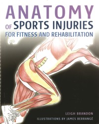
Anatomy of Sports Injuries offers a fascinating view inside the human body to help you understand the causes of the most common injuries that athletes and sports men and women will encounter, and the best ways to remedy them. With a comprehensive and up-to-date approach to injury rehabilitation, this book shows you how to find the cause of injuries, and not just treat the bit that hurts, aiding in the prevention of future injuries also. As with previous books in our anatomy series, this book will help you achieve better health through a better understanding of how your body works. Around 80 full-color anatomical illustrations introduce a variety of strength training exercises designed with common sports injuries in mind, showing the impact on the body of the exercises, including the muscles used and how they function together. The accompanying text helps the reader to replicate the exercises, describing the anatomical impact and explaining the benefits in the context of fitness in general, and in rehabilitating common injuries in particular.
Sie lesen das E-Book in den Legimi-Apps auf:
Seitenzahl: 182
Veröffentlichungsjahr: 2013
Das E-Book (TTS) können Sie hören im Abo „Legimi Premium” in Legimi-Apps auf:
Ähnliche


CONTENTS
PART 1OVERVIEW OF ANATOMY & INJURIES
How to use this book
Anatomical definitions and terminology
Systems of the body
Body planes and regions
Anatomical terms of position
Joint movements
Classification of Injuries
Injury Prevention
• Posture and muscle balance
• Event preparation
• Recovery
Long-term rehabilitation
• Tissue healing
• Healing phases
• Acute care
• Finding the cause of the injury
• Anatomy of the core
• Designing your programme
PART 2COMMON SPORTS INJURIES
Foot injuries
• Hallux valgus
• Metatarsal fracture
• Plantar fasciitis
Ankle and shin injuries
• Achilles tendonitis
• Achilles tendon rupture
• Ankle sprain
• Anterior compartment syndrome
• Shin splints
Knee injuries
• ACL sprain
• Baker’s cyst
• Chondromalacia patella
• Patella tendonitis
• Medial cartilage injury
• Medial collateral ligament sprain
• Osteoarthritis
• Posterior cruciate ligament sprain
• Quadriceps tendonitis
Thigh injuries
• Hamstring origin tendinopathy
• hamstring strain
• myositis ossificans
• quadriceps contusion
• quadriceps strain
• stress fracture of the femur
Groin injuries
• Gilmore’s groin
• Groin strain
• Inguinal hernia
• Osteitis pubis
Gluteal injuries
• hip bursitis
• piriformis syndrome
• sacroiliac joint dysfunction
• sciatica
Lumbar spine injuries
• Facet joint pain
• disc derangement
• spondylolysis and spondylolysthesis
Cervical and thoracic spine injuries
• Ankylosing spondylitis
• Atlas Subluxation Complex
• Scheuermann’s disease
• whiplash
Chest and abdominal injuries
• Abdominal hernia
• costochondritis
• pectoralis major strain
• rib fracture
• SC joint sprain
Shoulder injuries
• AC joint sprain
• biceps brachii strain
• dislocation
• fracture of the clavicle
• frozen shoulder
• glenoid labrum tear
• impingement syndrome
• rotator cuff strain
Elbow injuries
• Golfer’s elbow
• median nerve entrapment
• radial nerve entrapment
• tennis elbow
Wrist injuries
• Carpal tunnel syndrome
• fractured scaphoid
Environmental Injuries
• Dehydration
• frost bite
• heat stroke
• hypothermia
• sun burn
PART 3REHABILITATION
MOBILIZATIONS AND STRETCHES
• Mobilizations
• McKenzie push-up
• Median nerve mobilization
• Radial nerve mobilization
• Shoulder mobilization
Stretches
• Abdominals
• Adductors
• Anterior tibialis
• Calf
• Hamstrings
• Neck extensor
• Pectoralis minor
• Quadriceps
• Tensor fascia lata
EXERCISES
Postural exercises
• Hip and back extension
• Prone cobra
Stability exercises
• Four-point tummy vacuum
• Cable rotator cuff
• Cross band walking
• Deep cervical flexors
• Goof ball neck exercises
• Horse stance vertical
• Lower abdominals
• Stand/squat on balance board
• Supine lateral ball roll
• Touch toe drill
Strength exercises
• Deadlift
• Lunge
• Medicine ball shoulder external rotators
• Medicine ball shoulder internal rotators
• Romanian deadlift
• Single arm cable push
• Single arm dumbbell shrugs
• Single arm cable pull
• Supine hip extension on ball with belt
• Water jogging
• Wood chop
• Wrist extensors
• Wrist flexors
Glossary
Index
References and Resources
PART 1 – OVERVIEW OFANATOMY AND INJURIES
HOW TO USE THIS BOOK
Anatomy of Sports Injuries for Fitness and Rehabilitation is a visual and textual analysis of common sports injuries and rehabilitation of those injuries through effective exercises. It is also a guide to how to do the exercises properly and when to seek professional help to overcome your sports injury.
The book has three distinct parts: the first is a basic introduction to anatomical definitions, terminology and an overview of sports injuries. It also includes guidelines on injury prevention, acute care and first aid, manual therapy, long-term rehabilitation and lifestyle considerations.
Part two is divided into 13 sections covering a region of the body and highlights some of the most common injuries for that region. Each section defines individual injuries and their potential causes, treatment plans and statistics. Up to three mobilizations, stretches and/or exercises that may be used to help rehabilitate the injury are suggested as part of the corrective exercise programme following the acute phase of the injury.
Note that an injury can have many different causes and should be assessed by a trained professional to find the underlying causes. Any muscle imbalances should be highlighted at this stage and proper corrective stretching and strengthening should be given based on this information.
Disclaimer: Many of the exercises have a degree of risk of injury if done without adequate instruction and supervision. We recommend that you have a thorough assessment with a CHEK Practitioner, physiotherapist, osteopath or chiropractor before undertaking any of the exercises, and that you seek qualified instruction if you are a complete beginner. This book does not constitute medical advice and the author and publisher cannot be held liable for any loss, injury or inconvenience sustained by anyone using this book or the information contained in it.
Without a thorough assessment, the likelihood of full rehabilitation is greatly reduced, therefore, the stretches and exercises recommended may not be applicable to all.
Part three is an exercise section – a ‘how-to’ guide to doing the exercises as well as a visual and technical exercise analysis describing which muscles are being used. The start and finish position are usually depicted and training tips may be included.
The adult human body has more than 600 muscles and 206 bones; in this bookemphasis is placed on about 92 muscles involved in movement and stabilization. Many of the smaller muscles, as well the deep, small muscles of the spine and muscles of the hands and feet are not given specific attention.
This book is designed to help you improve your understanding of sports injuries and to overcome them and get back to performing at your best without the worry of further or future injury. Before starting a rehabilitation programme, the reader is advised to fully understand what phase of recovery they are in and introduce the right treatments and exercises at the right time (explained in part one). For instance, if stretches and exercises are used in the acute phase, this may further damage tissues and make the injury worse. Therefore, it is advised that you work through the book in the order it was written. In Part one, you will understand the anatomical definitions and terminology used in the book as well as a basic understanding of injuries and rehabilitation strategies. In Part two you will learn about your injury, while in Part three you will learn how to perform the exercises and stretches.
Ultimately, the injured tissues need to be conditioned to take the rigours of your sport in all planes of motion. This is known as end-stage rehab. While it is beyond the scope of this book to teach you end-stage rehab, the reader is advised to receive professional advice on strength and conditioning or read Anatomy of Strength and Fitness Training for Speed and Sport by Leigh Brandon.
ANATOMICAL DEFINITIONS AND TERMINOLOGY
Anatomy has its own language and although technical, it is quite logical, originating from Latin and Greek root words that make it easier to learn and understand the names of muscles, bones and other anatomy parts.
Whether you are an athlete, a student, a physio-therapist, a strength and conditioning coach or a CHEK practitioner, using the correct words and terminology enables you to interact with other professionals and professional materials.
Like most medical terms, anatomical terms are made up of small word parts, known as combining forms that fit together to make the full term. These ‘combining forms’ comprise roots, prefixes and suffixes. Knowing the different word parts allows you to unravel the word. Most anatomical terms only contain two parts: either a prefix and root or a root and suffix.
For example, if you take the terms ‘subscapular’ and ‘suprascapular’; the root is ‘scapula’, more commonly known as the shoulder blade. ‘Supra’ means ‘above’, hence ‘suprascapula’ means something above the shoulder blade. On the other hand, ‘sub’ means ‘below’, indicating in this instance something below the shoulder blade.
Common prefixes, suffixes and roots of anatomical terms
Word root
Meaning
Example
Definition
abdomin
pertaining to the abdomen
abdominal muscle
major muscle group of the abdominal region
acro
extremity
acromion
protruding feature on the scapula bone
articul
pertaining to the joint
articular surface
joint surface
brachi
pertaining to the arm
brachialis
arm muscle
cerv
pertaining to the neck
cervical vertebrae
the neck region of the spine
crani
skull
cranium
bones forming the skull
glute
buttock
gluteus maximus
buttock muscle
lig
to tie, to bind
ligament
joins bone to bone
pector
chest region
pectoralis major
chest muscle
Word parts used as prefixes
ab-
away from, from, off
abduction
movement away from the midline
ad-
increase, adherence, toward
adduction
movement towards the midline
ante-, antero-
before, in front
anterior
front aspect of the body
bi-
two, double
biceps brachii
two-headed arm muscle
circum-
around
circumduction
circular movement of a limb
cleido-
the clavicle
sternocleiomastoid
muscle, inserts into clavicle
con-
with, together
concentric contraction
contraction in which muscle attachments move together
costo-
rib
costal cartilage
rib cartilage
cune-
wedge
cuneiform
wedge-shaped foot bone
de-
down from
depression
downward movement of the shoulder blades
dors-
back
dorsiflexion
movement of the top side of the foot towards the shin
ec-
away from
eccentric contractions
contraction in which muscle attachments move apart
epi-
upon
epicondyle
feature of a bone, located above a condyle
fasci-
band
tensor fasciae latae
small band-like muscle of the hip
flex-
bend
flexion
movement closing the angle of a joint
infra-
below, beneath
infraspinatus
muscle situated below the spine of the scapula
meta-
after, behind
metatarsals
bones of the foot, distal to the tarsals
post-
after, behind
posterior
rear aspect of the body
pron-
bent forward
prone position
lying face down
proximo-
nearest
proximal
nearest the root of a limb
quadr-
four
quadriceps
four-part muscle group on the anterior thigh
re-
back, again
retraction
pulling of the shoulder blades towards the midline
serrat-
saw
serratus anterior
muscle with a saw-like edge
sub-
beneath, inferior
subscapularis
muscle beneath the scapula
super, supra-
over, above, excessive
supraspinatus
muscle above the spine of the scapula
superior
toward the head
thoraco-
the chest, thorax
thoracic vertebrae
in the region of the thorax
trans-
across
transverse abdominus
muscle crossing the abdomen
tri-
three
triceps brachii
three-headed muscle of the upper arm
tuber-
swelling
tubercle
small rounded projection on a bone
Word parts used as suffixes
-al, ac
pertaining to
iliac crest
pertaining to the ilium
-cep
head
biceps brachii
two-headed arm muscle
-ic
pertaining to
thoracic vertebrae
pertaining to the thorax
-oid
like, in the shape of
rhomboid
upper back muscle, in the shape of a rhomboid
-phragm
partition
diaphragm
muscle separating the thorax and abdomen
SYSTEMS OF THE BODY
The human body can be viewed as an integration of approximately 12 distinct systems that continuously interact to control a multitude of complex functions. These systems are a co-ordinated assembly of organs, each with specific capabilities, whose tissue structures suit a similar purpose and function.
This book illustrates and analyzes the systems that control movement and posture, namely the muscular and skeletal systems; often referred to jointly as the musculoskeletal system.
The other systems are the cardiovascular, lymphatic, nervous, endocrine, integumentary, respiratory, digestive, urinary, immune and reproductive systems.
The muscular system
The muscular system facilitates movement, maintenance of posture and the production of heat and energy. It is made up of three types of muscle tissue: cardiac, smooth and striated.
Cardiac muscle forms the walls in the heart, while smooth muscles tissue is found in the walls of internal organs such as the stomach and blood vessels. Both are activated involuntarily via the autonomic nervous system and hormonal action.
Striated muscle makes up the bulk of the muscles as we commonly know them. The skeletal system includes the tendons that attach muscle to bone, as well as the connective tissue that surrounds the muscle tissue, which is called fascia.
A human male weighing 70 kg (154 lbs) has approximately 25–35 kg (55–77 lbs) of skeletal tissue.
Muscle attachments
Muscles attach to bone via tendons. The attachment points are referred to as the origin and the insertion.
The origin is the point of attachment that is proximal (closest to the root of a limb) or closest to the midline, or centre of the body. It is usually the least moveable point, acting as the anchor in muscle contraction.
The insertion is the point of attachment that is distal (furthest from the root of a limb) or furthest from the midline or centre of the body. The insertion is usually the most moveable part, and can be drawn towards the origin.
Knowing the origin and insertion of a muscle, which joint or joints the muscle crosses and what movement is caused at that joint or joints is a key element of exercise analysis.
There are typical features on all bones that act as convenient attachment points for the muscles. A description of typical bone features is given in the table on page 11.
Typical features on a bone
Feature
Description
Examples
Condyle
Large, rounded projection at a joint, that usually articulates with another bone
Medial and lateral condyle of the femur Lateral condyle of the tibia
Epicondyle
Projection located above the condyle
Medial or lateral epicondyle of the humerus
Facet
Small, flat joint surfaces
Facet joints of the vertebrae
Head
Significant, rounded projection at the proximal end of a bone, usually forming a joint
Head of the humerus
Crest
Ridge-like, narrow projection
Iliac crest of the pelvis
Line, Linea
Lesser significant ridge, running along a bone
Linea aspera of the femur
Process
Any significant projection
Coracoid and acromion process of the scapula Olecranon process of the ulna at the elbow joint
Spine, Spinous process
Significant, slender projection away from the surface of the bone
Spinous processes of the vertebrae Spine of the scapula
Suture
Joint line between two bones forming a fixed or semi-fixed joint
Sutures that join the bones of the skull
Trochanter
Very large projection
Greater trochanter of the femur
Tubercle
Small, rounded projection
Greater tubercles of the humerus
Tuberosity
Large, rounded or roughened projection
Ischial tuberosities on the pelvis
Foramen
Rounded hole or opening in a bone
The vertebral foramen running down the length of the spine, in which the spinal cord is housed
Fossa
Hollow, shallow or flattened surface on a bone
Supraspinous and infraspinous fossa on the scapula
The word ‘skeleton’ originates from a Greek word meaning ‘dried up’. Infants are born with about 350 bones, many of which fuse as they grow, forming single bones, resulting in the 206 bones that an adult has.
The skeletal system
This consists of bones, ligaments (which join bone to bone) and joints. Joints are referred to as articulations and are sometimes classified as a separate system, the articular system.
Apart from facilitating movement, the primary functions of the skeletal system include supporting the muscles, protecting the soft tissues and internal organs, the storage of surplus minerals and the formation of red blood cells in the bone marrow of the long bones.
Integrated systems
The body’s systems are completely and intricately interdependent. For movement to take place, for example, the respiratory system brings in oxygen and the digestive system breaks down our food into essential nutrients. The cardiovascular system then carries the oxygen and nutrients to the working muscles via the blood to facilitate the energy reactions that result in physical work being done.
The lymphatic and circulatory systems help to carry away the waste products of these energy reactions, which are later converted and/or excreted by the digestive and urinary systems. The nervous system interacts with the muscles to facilitate the contraction and relaxation of the muscle tissue. The articular system of joints allows the levers of the body to move.
The femur (thigh bone) is about one quarter of a person’s height. It is also the largest, heaviest and strongest bone in the body. The shortest bone, the stirrup bone in the ear is only about 2.5 mm long. An adult’s skeleton weights about 9 kg (20 lb)
BODY PLANES AND REGIONS
When learning anatomy and analyzing movement, we refer to a standard reference position of the human body, known as the anatomical position (see illustrations below). All movements and locations of anatomical structures are named as if the person were standing in this position.
Regional anatomy
This book is a technical labelling guide to the different superficial parts of the body. In anatomical language, common names such as ‘head’ are replaced with anatomical terms derived from Latin, such as ‘cranial’ or ‘cranium’.
Within the different body regions there are sub-regions. For example, within the cranial region are the frontal, occipital, parietal and temporal sub-regions.
Anatomical planes
The body can be divided into three imaginary planes of reference, each perpendicular to the other.
The sagittal plane passes through the body from front to back, dividing it into a right half and a left half. The midline of the body is called the median. If the body is divided in the sagittal plane, directly through its median, this is known as the median sagittal plane. The coronal (frontal plane) passes through the body from top to bottom, dividing it into front and back sections.
The transverse (horizontal) plane passes through the middle of the body at right angles, dividing it into a top and a bottom section.
An anatomical cross-section of the internal structures of the body can be viewed in any one of these planes, which are also described as ‘planes of motion’, as the joint movements are defined in relation to one of the three planes. Understanding into which plane an anatomical cross-section is divided will help you know what you are looking at and from which viewpoint.
ANATOMICAL TERMS
There are standard anatomical terms that describe the position or direction of one structure of the body and its relationship to other structures or parts of the body.
The human body is a complex, three dimensional structure. Knowing the proper anatomical terms of position and direction will help you to compare one point on the body with another and understand where it is situated in relation to the other anatomical features.
These terms are standard, no matter whether the person is standing, seated or lying, and are named as if the person was standing in the anatomical position (see page xx). The terms of direction should not be confused with joint movements (see pages 17–20).
Anatomical terms of position and direction
Position
Definition
Example of usage
Anterior
Towards the front, pertaining to the front
The pectoral muscles are found on the anterior aspect of the body
Posterior
Towards the back, pertaining to the back
The calf muscles are situated on the posterior surface of the lower leg
Superior
Above another structure, towards the head
The knee is superior to the ankle
Inferior
Below another structure, towards the feet
The hip is inferior to the shoulder
Lateral
Away from the midline, on or towards the outside
The radial bone is lateral to the ulna
Medial
Towards the midline, pertaining to the middle or centre
The tibial bone is medial to the fibula
Proximal
Closest to the trunk or root of a limb; sometimes used to refer to the origin of a muscle
The shoulder joint is proximal to the elbow
Distal
Situated away from the midline or centre of the body, or root of a limb; sometimes used to refer to a point away from the origin of a muscle
The knee joint is distal to the hip
Superficial
Closer to the surface of the body, more towards the surface of the body than another structure
The rectus abdominus is the most superficial muscle of the abdominal wall
Deep
Further from the surface, relatively deeper into the body than another structure
The transverse abdominus is the deepest muscle of the abdominal wall
Prone
Lying face down
A prone cobra exercise is performed from a lying start position
Supine
Lying on the back, face upwards
A bench press exercise is performed from a supine position
JOINT MOVEMENTS
Knowing and understanding movement (which joint is moving and how it moves) is essential in order to analyze a complex exercise. This book has done the job of joint identification for you, and understanding this section will help improve your exercise analysis.
Types of joints
Some joints are fixed or semi-fixed, allowing little or no movement. For instance, the bones of the skull join together in structures known as sutures to form fixed joints; but where the spine joins the pelvis, the sacroiliac (‘sacro’ from sacrum and ‘iliac’ pertaining to the pelvic crest) joint is semi-fixed and allows minimal movement.
A third category called synovial joints are free-moving and move in different ways determined by their particular shape, size and structure.
Synovial joints are the most common joints in the body. They are categorized by a joint capsule that surrounds the articulation, the inner membrane of which secretes lubricating synovial fluid, stimulated by movement. Typical synovial joints include the shoulder, knee, hip, ankle, joints of the feet and hands and the vertebral joints.
Joint action
When performing an activity such as lifting weights or running, the combination of nerve stimulation and muscular contraction facilitates the movement that occurs at the synovial joints.


