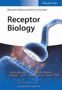
69,99 €
Mehr erfahren.
- Herausgeber: Wiley-VCH
- Kategorie: Wissenschaft und neue Technologien
- Sprache: Englisch
This book is geared to every student in biology, pharmacy and medicine who needs to become familiar with receptor mediated signaling. The text starts with explaining some basics in membrane biochemistry, hormone biology and the concept of receptor based signaling as the main form of communication between cells and of cells with the environment. It goes on covering each receptor superfamily in detail including their structure and evolutionary context. The last part focusses exclusively on examples where thorough knowledge of receptors is critical: pharmaceutical research, developmental biology, neurobiology and evolutionary biology. Richly illustrated, the book is perfectly suited for all courses covering receptor based signaling, regardless whether they are part of the biology, medicine or pharmacology program.
Sie lesen das E-Book in den Legimi-Apps auf:
Seitenzahl: 694
Veröffentlichungsjahr: 2016
Ähnliche


Table of Contents
Cover
Title Page
Copyright
Dedication
Acknowledgments
Part I: Introduction
Chapter 1: Introduction
1.1 Receptors and Signaling
1.2 Types of Receptors and Hormones
1.3 Receptors Are the Chemical Expression of Reality
Chapter 2: The Origins of Chemical Thinking
2.1 Overview of Early Pharmacological History
2.2 Modern Pharmacology
2.3 Phylogenetics of Signaling
Part II: Fundamentals
Chapter 3: Membranes and Proteins
3.1 Membranes
3.2 The Nature and Function of Proteins
Chapter 4: Hormones as First Messengers
4.1 Hormones and Cellular Communication
4.2 Types of Hormones
4.3 Vertebrate Hormones and Transmitters
4.4 Analgesics and Venoms as Receptor Ligands
Chapter 5: Receptor Theory
5.1 The Materialization of Receptors
5.2 Receptor Mechanisms
5.3 Binding Theory
5.4 Visualizing Receptor Structure and Function
5.5 Proteomics Approaches to Receptor Efficacy
5.6 Physical Factors Affecting Receptor Binding
Part III: Receptor Types and Function
Chapter 6: Transduction I: Ion Channels and Transporters
6.1 Introduction
6.2 Small Molecule Channels
6.3 Transporters
6.4 Major Intrinsic Proteins
6.5 Ligand-Gated Ion Channels
Chapter 7: Transduction II: G-Protein-Coupled Receptors
7.1 Introduction
7.2 Families of G-Protein-Coupled Receptors
7.3 Transduction Mechanisms
7.4 The Major Families of G Protein-Coupled Receptors
Chapter 8: Transduction III: Receptor Kinases and Immunoglobulins
8.1 Protein Kinases
8.2 Receptors for Cell Division and Metabolism
8.3 Receptor Serine/Threonine Kinases
8.4 The Guanylyl Cyclase Receptor Subfamily – Natriuretic Peptide Receptors
8.5 Non-Kinase Molecules – LDL Receptors
8.6 Cell–Cell Contact Signaling
8.7 Immune System Receptors, Antibodies, and Cytokines
Chapter 9: Transduction IV: Nuclear Receptors
9.1 Introduction
9.2 Genomic Actions of Nuclear Receptors
9.3 Actions of Receptor Antagonists
9.4 Non-Traditional Actions of Steroid-Like Hormones and Their Receptors
Part IV: Applications
Chapter 10: Signaling Complexity
10.1 Introduction
10.2 Experimental Determination of Signaling Cascades
10.3 Transduction across the Membrane
10.4 Complexity in Cross Talk – Roles of PIP3, Akt, and PDK1
10.5 Role in Cancer
10.6 Signaling Mediated by Gas Molecules
Chapter 11: Cellular Interactions in Development
11.1 Introduction
11.2 The Origins of Multicellularity
11.3 The Origin of Symmetry and Axes
11.4 Fertilization and Organization of the Multicellular Body Plan
11.5 Differentiation of Triploblastic Embryos – Organogenesis
11.6 Programmed Cell Death (Apoptosis)
Chapter 12: Receptor Mechanisms in Disease Processes
12.1 Genetic Basis for Receptor Function
12.2 Receptor Pathologies
12.3 Signaling Mutations Leading to Cancer
Chapter 13: Receptors and the Mind
13.1 Origins of Behavior
13.2 Nervous Systems
13.3 Animal Memory: Invertebrates
13.4 Animal Memory: Vertebrates
13.5 Receptors and Behavior: Addiction, Tolerance, and Dependence
Chapter 14: Evolution of Receptors, Transmitters, and Hormones
14.1 Introduction
14.2 Origins of Transmitters and Receptors
14.3 Evolution of Hormones
14.4 Evolution of Receptor Superfamilies
14.5 Evolution of Receptor Antagonism
14.6 A Final Note
Glossary
References
Index
End User License Agreement
Pages
xiii
1
3
4
5
6
7
9
10
11
12
13
14
15
17
18
19
20
21
22
23
24
25
26
27
28
29
30
31
32
33
34
35
36
37
38
39
40
41
42
43
44
45
46
47
48
49
50
51
52
53
54
55
56
57
58
59
60
61
63
65
66
67
68
69
70
71
72
73
74
75
76
77
78
79
80
81
82
83
85
86
87
88
89
90
91
92
93
94
95
96
97
98
99
100
101
102
103
104
105
106
107
108
109
110
111
112
113
114
115
116
117
118
119
121
122
123
124
125
126
127
128
129
130
131
132
133
135
136
137
138
139
140
141
142
143
144
145
146
147
148
149
150
151
152
153
154
155
156
157
158
159
160
161
162
163
164
165
166
167
169
170
171
172
173
174
175
176
177
178
179
180
181
182
183
184
185
186
187
188
189
190
191
192
193
194
195
197
198
199
200
201
202
203
204
205
206
207
208
209
210
211
212
213
214
227
228
229
230
231
232
233
234
235
236
237
238
239
241
242
243
244
245
246
247
248
249
250
Guide
Cover
Table of Contents
Part I: Introduction
Begin Reading
List of Illustrations
Chapter 1: Introduction
Figure 1.1 Overview of the four major receptor types. (A) Ion channel with extracellular
domain
labeled in red and transmembrane chains labeled in green; (B) G-protein-coupled receptor with extracellular domains labeled in red, seven transmembrane domains labeled in green; (C) receptor tyrosine kinase with transmembrane domain labeled in green, extracellular regions in red, and intracellular regions in blue. Black lines represent lipid bilayer; and (D) nucleus (dashed line) with nuclear receptors labeled in blue dimerizing on a DNA template labeled in black.
Chapter 2: The Origins of Chemical Thinking
Figure 2.1 (a) Axonal ending of a neuron at a postsynaptic cell (nerve or skeletal muscle). The axon is not directly connected to the muscle cell, but is separated by a gap or synapse. The transmitter (green spheres) is synthesized in the ending within vesicles; it is released in response to an action potential. The transmitter diffuses to receptors on the postsynaptic cell. (b) An antagonist such as curare (red ovals) binds to the same receptor at the active site, preventing the transmitter from binding and thereby preventing the effects of nerve stimulation. http://kin450-neurophysiology.wikispaces.com/Synaptic+Transmission. Accessed 7 February 2015.
Figure 2.2 Ehrlich's representation of “side chains” representing his receptor concept. The fish-shaped objects binding to the chains are toxins or immunologic antigens. Here he shows a cell surface with a “side chain” projecting off the cell. This chain has a specific binding site for a toxin or antigen.
Figure 2.3 (a): Cross section of the spinal cord in the chest region; a sympathetic preganglionic nerve exits and synapses in the ganglion at (b) with a postganglionic neuron. Langley discovered the synapse at (b) by cutting the presynaptic nerve between (a) and (b), then adding nicotine to the ganglion.
Figure 2.4 Phylogenetic diagram of the three domains of living organisms, with overall indication of the receptor types present in each and some functions of each. LUCA: “last universal common ancestor,” the hypothesized cellular ancestor of living organisms. The Eukarya and Archaea are more closely related to each other than either is to the bacteria.
Chapter 3: Membranes and Proteins
Figure 3.1 The fluid mosaic model of the cell membrane. The characteristics of this model are the phospholipid bilayer and the presence of globular proteins (large molecules) within and on the surface of the membrane. The portions of the proteins facing the extracellular and cytoplasmic solutions are hydrophilic; central parts are hydrophobic. Cholesterol molecules within the membrane are not shown here. https://s3.amazonaws.com/engrade-myfiles/4034298582543629/fluidmosaic.jpg. Accessed 7 February 2015.
Figure 3.2 Spontaneous arrangements of phospholipids on water surface. (a) Monolayer of lipid on water surface in contact with air. (b) Lipid bilayer within the water environment.
Figure 3.3 Danielli–Davson model of membrane lipids and proteins. The proteins (blue) were thought to be flat sheets lying on the lipid bilayer inside and outside the cell.
Figure 3.4 Molecular structures of (a) sphingomyelin, a
sphingolipid
and (b) phosphatidylcholine, a glycerol-based
phospholipid.
Both have phosphatidyl portions. The dotted squares enclose the head of the sphingosine group (a) and the glycerol group (b). Each vertex of the jagged lines represents a CH
2
unit of the fatty acid.
Figure 3.5 Molecular structure of cholesterol.
Figure 3.6 Post-translation processing of proteins. Golgi apparatus membranes shown at left.
a
(blue): Secreted protein, for example, a peptide hormone. Because most of the peptide is hydrophilic, it crosses the ER membrane and resides in the cisterna of the ER. The vesicle containing the peptide fuses with the cell membrane and the peptide finds itself outside the cell.
b
(red): Membrane protein, for example, a receptor. The signal peptide promotes uptake of the growing chain, but because of hydrophobic regions, it remains in the Golgi membrane. As the vesicle fuses with the cell membrane, the receptor remains part of the membrane.
Figure 3.7 Linear and globular proteins. (a) Three linear peptide strands of collagen (red, blue, and green). (b) Four globular peptide strands of hemoglobin (blue and green), each with iron atom bound to heme (red).
Figure 3.8 Formation of the peptide bond in proteins when the terminal hydroxyl from one amino acid combines with the initial hydrogen of another.
Figure 3.9 Table of polarities of amino acids, in order of hydropathy score (positive = nonpolar).
Figure 3.10 Relationship between receptor protein hydropathy plot and orientation within the membrane. (a) Plot of hydropathy values (+ represents hydrophobic amino acids) for rhodopsin (about 350 amino acids). I–VII: membrane domains. (b) Probable orientation of the seven transmembrane (TM) domains within the membrane. The entire molecule forms a roughly circular structure rather than a linear structure.
Figure 3.11 Alpha-helix structure of proteins. (a, b) The shape of the helix is stabilized by hydrogen bonds between adjacent turns of the helix. This structure is found in most proteins, but is especially important in membrane proteins as the transmembrane domain (Chapters 6–8).
Figure 3.12 Pleated sheet structure of proteins. (a, b) Hydrogen bonds form between the CO of one strand to the NH of the parallel strand. This structure is seen in many proteins, but is often a part of the extracellular binding domain of ligand-gated ion channels (Chapter 6).
Chapter 4: Hormones as First Messengers
Figure 4.1 Simplified phylogeny of life, showing the five major eukaryotic groups – unikonta, excavata, archaeplastida, rhizaria, and chromalveolata. Organisms mentioned in this chapter are indicated. 1°: Primary endosymbiosis of mitochondrion (all eukaryotes) and chloroplast (line leading to archaeplastida. 2°: Two separate endosymbiotic events in which a eukaryotic green alga was incorporated into an early excavate and a rhizaria; also, a eukaryotic red alga was incorporated into an early brown alga (kelp).
Figure 4.2 Quorum sensing in bacteria. (a) Gene Lux1 encodes an enzyme that synthesizes autoinducer (pentagon), which diffuses away from the cell. If enough cells are in the vicinity, large amounts of autoinducer are produced, and diffuse into nearby cells. There, they bind to transcription factor LuxR and activate target genes. (b) Structure of the autoinducer AI-1. (c) Structure of the autoinducer AI-2.
Figure 4.3 Molecular structures of chromalveolate signaling molecules. (a) The monosaccharide mannose. (b) The aldehyde (2E, 4E), decadienal. (c) The aromatic hydrocarbon lamoxirene. (d) The hydrocarbon fucoserratene.
Figure 4.4 Molecular structures of signaling molecules for amoebozoa aggregation and differentiation. (a) cAMP and (b) glorin, a hydrocarbon and peptide-containing signaling molecule. The red oval encloses glutamic acid and the green oval encloses ornithine.
Figure 4.5 (a) Amino acid sequence of yeast-mating-type peptide MAT-alpha. (b) Sequence of yeast-mating-type peptide MAT-a. MAT-a can have either a valine or a leucine at position 6.
Figure 4.6 Molecular structure of bombykol, a volatile long chain alcohol used by female silkworm moths to attract males.
Figure 4.7 Posterior pituitary structure and locations of hypothalamic neurosecretory cells that release its hormones. Axons of neurosecretory cells extend from the hypothalamus to the posterior pituitary.
Figure 4.8 Amino acid sequences of the posterior pituitary hormones, (a) oxytocin and (b) vasopressin. The two-dimensional structures are very similar, but the two different amino acids (in circles) determine the different functions of each.
Figure 4.9 Structure of the anterior pituitary and its relation to hypothalamic releasing hormones. Axons of neurosecretory cells stop at portal vessels at the base of the hypothalamus.
Figure 4.10 Post-translation processing of the pituitary peptide pro-opiomelanocortin. MSH: melanocyte-stimulating hormone and CLIP: corticotropin-like intermediate peptide.
Figure 4.11 Amino acid sequences of the gastrointestinal peptide hormones cholecystokinin (CCK) and gastrin. Underlining shows a short section of sequence identity.
Figure 4.12 Feedback loops in production of CCK, secretin, gastrin, and gastric inhibitory peptide (GIP).
Figure 4.13 Amino acid sequences of endothelin-1 and sarafotoxin-68, showing areas of sequence identity and disulfide bond locations.
Figure 4.14 Synthetic pathway for catecholamine neurotransmitters and hormones. Dopamine, norepinephrine, and epinephrine serve as neurotransmitters.
Figure 4.15 Molecular structures of (a) acetylcholine, (b) histamine, (c) serotonin, and (d) melatonin.
Figure 4.16 Molecular structures of nucleotide base alone (a), nucleoside (base plus ribose sugar – b), and nucleotide monophosphate (base plus sugar plus phosphate – c). These illustrations refer to the base adenine; similar molecules form for the other bases, guanine, cytosine, and uracil, and are based on the RNA version of each nucleotide.
Figure 4.17 Synthesis of prostaglandins and related signaling compounds. COX: cyclooxygenase; terms with PG refer to prostaglandins; PGI
2
is also called
prostacyclin
; and TX refers to thromboxane.
Figure 4.18 (a) Structure of arachidonic acid, from which the prostaglandins are synthesized by the action of cyclooxygenase. (b) Structure of an example of prostaglandins, PGD
2
. The red lines represent equivalent carbon–carbon bonds, and in all PGs an oxygenated cyclic structure is formed between carbons 8 and 12. (c) Thromboxane A
2
(TXA
2
), with cyclic oxygen on ring. (d) Prostacyclin (PGI
2
), with cyclic oxygen between ring and chain.
Figure 4.19 Molecular structures of cannabinoids. (a) Anandamide, an endocannabinoid. (b) Δ
9
-Tetrahydrocannabinol, the active ingredient of the marijuana plant.
Figure 4.20 Molecular structures of amino acid transmitters. (a) Glutamic acid. (b) Gamma-amino butyric acid. (c) Glycine.
Figure 4.21 Overall structure and disulfide bonds of insulin. Proinsulin is synthesized as the red and black sequence. Cleavage of the amino acids at the black and red junctions results in the release of the nonfunctional C peptide (red); disulfide bonds connect the two short strands (black) that form the functional insulin.
Figure 4.22 Molecular structures of hormones for nuclear receptors. Above: family 3 receptor ligands; below: family 1 receptor ligands. Receptor families are described in Chapter 9.
Figure 4.23 Synthesis of the major steroid hormones. Estrogen is the final steroid in the sex steroid pathway; its conversion from testosterone is catalyzed by the enzyme aromatase.
Figure 4.24 In different tissues of the male mammal, testosterone is either the source of the differentiating signal for male differentiation or the substrate for enzymes converting it to dihydrotestosterone or estrogen (red underlining). All three steroids have roles in male development.
Chapter 5: Receptor Theory
Figure 5.1 Top view of the nicotinic ACh receptor, showing the five peptide subunits (2α, β, δ, ϵ) in a circular arrangement around the central ion channel. Two ACh molecules bind, one to the α–δ pair (green and blue at left) and the other to the α–ϵ pair (green and orange at right). Binding is of three types: A (left): Ionic bonds between the quaternary N of the ACh and a negative charge on the δ and ϵ subunits. B (right): Hydrogen bonds between the carboxyl O of the ACh and an H on the α subunits. C (right): van der Waals bonds may also form between the intermediate portions of the ACh and nonionic amino acids of the receptor subunits. Only the β subunit seems not to be involved in specific bonding.
Figure 5.2 Graph of an experiment in which the amount of ligand bound to receptor (AR) is illustrated as a function of the ligand concentration. The
K
d
is about 2 nM. Agonist (ligand) concentration has a linear axis.
Figure 5.3 Apparatus for recording smooth muscle contractions. The tissue is within an organ bath at right, inside a larger bath that maintains temperature. Isometric tension is shown recorded at top right; increases in tension are caused by increasing concentrations of agonist, which are added to the organ bath in stepwise fashion. Tissue can be washed free of agonist by allowing solution (upper left) to replace organ bath solution. Stirring apparatus at left mixes large chamber.
Figure 5.4 Response of tissue to hormone or neurotransmitter (agonist). Data from Clark [123] for action of ACh on frog rectus abdominis muscle. Same data plotted on (a) linear
X
-axis and (b) log
X
axis.
Figure 5.5 Clark's model of the relation between receptor occupancy (
X
axis) and response (
Y
axis). A: Linear relation between binding and response such that full response is reached only at full occupancy. C: The relation for a competitive antagonist, which may bind 100% of receptors without initiating a response.
Figure 5.6 Early model of the relation between receptor occupancy (
X
axis) and response (
Y
axis). Partial agonists: Linear relation between binding and response (proposed by Clark) such that full response is reached only at full occupancy. Full agonists: significant response is reached when only a small proportion of receptors are bound. Competitive antagonists, which may bind 100% of receptors without initiating a response. Partial agonists may be defined as agonists with response patterns between 0 and the linear relation; full agonists by strongly hyperbolic response patterns.
Figure 5.7 Del Castillo and Katz model for ion channel agonist binding and activation.
K
is the affinity of agonist binding.
E
is the efficacy of channel activation. This hypothesis shows the complex relationship between occupation and action, as inactive receptors may be vacant or occupied, and occupied receptors might be active or inactive. The portion within the ellipse is the occupied receptor converting from the inactive state to the active state.
Figure 5.8 Two aspects of the two-state model of receptor activation. The boxed series represents classical hypotheses of affinity (
K
A
) and efficacy (
E
1
) according to the occupancy model and the Del Castillo and Katz [128] model. The remainder illustrates alterations accounting for active unbound receptors (
E
0
) and conformational change of receptor upon agonist binding (
K
A*
).
Figure 5.9 Ternary complex model. At top left, the agonist, receptor, and G protein are unbound. At top right, the agonist binds to receptor; at bottom right the G protein binds the others; at bottom left, the agonist dissociates. When initial state is reached again (top left); the receptor and G protein dissociate. The original Figure of Ref. [129] used the letter X for what we now know is the G protein; it also illustrated coupling of the ARG to the effector, adenylyl cyclase.
Figure 5.10 Extended ternary complex model. The four interactions at left form the ternary complex model (Figure 5.9); the extension involves incorporating the G-protein binding process along with its activating components (AR*G and R*G) at right. The R*G is not predicted to occur in the ternary complex model.
Figure 5.11 GTPase activity in isolated cell membranes in response to different ligands. Data show that receptor activity can be non-zero in the absence of agonist binding, and that different “agonists” have different modes of interacting with receptor. The neutral agonist in this model is equivalent to the competitive inhibitors described earlier; an inverse agonist creates a new type of inhibitory interaction.
Figure 5.12 (a)
S
-(−)-3-(3-hydroxyphenyl)-
N
-propylpiperidine (
S
-3-PPP) binds to the dopamine D2 receptor to activate G
o
, as well as to prevent G
i
activation (red line indicates inverse agonism). (b) n-Propyl norapomorphine (NPA) binds to the same receptor, acting as an agonist of both G
o
and G
i
.
Figure 5.13 Cubic ternary complex model of G-protein-coupled receptor interactions. R: inactive receptor; R*: active receptor; A: agonist; and G: G protein. The front face and red arrows represent the extended ternary complex model from Figure 5.10. In the cubic ternary model, all states on the right side of the cube are potentially “active.”
Figure 5.14 Explanation of the different states of agonism and antagonism. Receptor alters between R and R* states, and different types of agonists have different affinities for one or the other of those states. The thickness of the arrow is proportional to agonist affinity for the indicated state, leading to each circled agonist or antagonist description. Protean agonism is not shown, but would be the ability of a given agonist to be either an inverse or partial agonist on the same receptor.
Figure 5.15 Schild analysis for the response of rat trachea to carbachol; competitive antagonist – pirenzepine. (a) Dose–response curves in the absence (open circles) and in the presence of pirenzepine from 300 nM to 10 μM. The antagonist causes concentration-dependent rightward shifts in the dose–response curve. (b) Use of Equation (5.8) to graph the Schild regression that allows calculation of pA
2
.
Y
axis: log (dose ratio – 1) and
X
-axis: log [pirenzepine]. In this case, PA2 is approximately −6.9. Dotted line: linear best fit; solid line: line with slope = 1.
Figure 5.16 Radiolabeled compounds used for radioligand studies. (a)
125
I-labeled tyrosine as part of the insulin molecule. (b)
3
H-labeled dihydro-alprenolol, a beta-adrenoceptor antagonist.
Figure 5.17 (a) Total and non-specific binding for an equilibrium binding experiment. (b) Specific binding (difference between total and non-specific binding). Both Figure use arbitrary units of binding and concentration.
Figure 5.18 Competition study to measure
K
i
of antagonists in bovine tracheal smooth muscle. The vertical axis shows binding of a radiolabeled substance that binds to all types of muscarinic cholinergic receptors. The compounds listed in the legend are competitive antagonists that bind specifically to different types of muscarinic receptors.
Figure 5.19 Interference patterns in an inorganic crystal, illustrating how constructive and destructive patterns arise from incident X-rays, allowing estimate of the atomic structure of the crystal. Structures of organic crystals are more complex, and are more difficult to determine. Panels (a, b) from http://en.wikipedia.org/wiki/Bragg's_law.
Chapter 6: Transduction I: Ion Channels and Transporters
Figure 6.1 Mechanosensitive channel from
Mycobacterium.
(a) The monomer polypeptide consists of two transmembrane (TM) regions shown in blue and one cytoplasmic region in red. (b) The full channel protein is homopentameric, shown in shades of blue and red to contrast the individual monomers. When membrane is unstretched (b), channel is not open. (c) Top-down orientation of (b). (d) Membrane stretch acts in a rotational fashion to open the channel to 3 nm, allowing passage of ions or other osmotically important molecules. Images were created using Rasmol [8] from PDB ID 2OAR [156].
Figure 6.2 Example of recording of action potential from single neuron of squid giant axon. Rise in trace indicates opening of voltage-gated Na
+
channels.
Figure 6.3 Patch clamp method and application. (a) Cell with microelectrode applied to surface. Suction produces high-resistance seal, and further pulling generates electrode contact with cell interior or patch. (b) Current recordings from a single patch that contains two channels. At each depolarization, one or two channels open. (c) An average of opening currents from many applications of 40 mV stimuli on the same membrane patch.
Figure 6.4 Diagram of potassium channel families and subfamilies. Three structural classes are shown defined by their subunit structure, having six (6TM), four (4TM), or two (2TM) transmembrane segments. The KcsA and GIRK channels consist of tetramers of 2TM channels. The 4TM channels control resting neuronal potentials. The colored TM regions at left represent a 4TM structure containing the
voltage-sensing domain
. The Figure at left also show combinations of 4TM and 2TM domains to construct the large family of 6TM channels, only some of which are shown. For clarity, representatives of the 4TM and 2TM structural class are not shown.
Figure 6.5 The voltage-gated K
+
channel. The front and rear 6TM domains have been removed. Two K
+
ions are visible in the channel, and the voltage sensors are shown in red. Modified from Ref. [170]. Used with permission.
Figure 6.6 KcsA channel – organization. Each of the four peptides is shown in a different color. The selectivity filter is shown in the center as the thin bent domains of the peptides (all four peptides contribute to the filter, but only two of the peptides are visible in this diagram).
Figure 6.7 The GIRK channel, a tetrameric 2TM K
+
channel. (a) Graph of current (
Y
axis) and voltage (
X-
axis) relations of two types of K
+
channel; the linear channel remains open at negative and positive voltages; the inward rectifying channel closes at positive voltages. (b) Architecture of the channel (red) in the membrane; also shown is GPCR (e.g., muscarinic receptor) that activates the GIRK. The Gβγ protein binds to the channel following binding of ACh to receptor.
Figure 6.8 The bacterial voltage-gated Na
+
channel. The channel consists of four identical polypeptides, each with 6TM domains. Here, three of the four peptides are shown, and one is shown in color; TM domains 1–6 are numbered. The two helices of the pore loop (L) are indicated between TM5 and 6.
Figure 6.9 The vertebrate voltage-gated Na channel. Membrane architecture, showing its three proteins: α chain, β
1
chain, and β
2
chain.
Figure 6.10 The voltage-gated Ca channel. It has five different peptides: α
1
, α
2
, β, γ, and δ (γ subunits are not shown). Cylinders represent TM domains; plus signs represent TM domains that form the pore and selectivity filter.
Figure 6.11 Changes in membrane potential and tension of a ventricular cell over a single heartbeat. Action potential (short Na
+
channel opening) initiates Ca
2+
channel opening, which lasts 100 ms. The Na
+
channels cannot reopen while the Ca
2+
channel is open, so the cell is refractory during that period.
Figure 6.12 Generalized structure of members of the TRP Ca
2+
channel family. Both N- and C-termini are in the cytoplasm. Six TM domains (S1–S6) form the channel. A pore-forming loop (P) lies between S5 and S6. Residues near the C-terminus may be involved in the binding of phosphatidyl inositol phosphate, a Ca
2+
channel regulator.
Figure 6.13 Functions of TRPM channel in taste transduction. Binding of sweet, bitter, or umami taste stimulus (diamond) to G-protein-coupled taste receptors (R) – discussed in Chapter 7 – leads to release of βγ subunits of the G protein, which activates PLCβ2, leading to IP
3
release. IP
3
activates its receptors (IP
3
R), which release Ca
2+
from intracellular stores. The Ca
2+
opens TRPM5 channels, leading to an influx of Na
+
and depolarization of the cell.
Figure 6.14 Model of the glutamate transporter. The molecule forms a large basin in the extracellular side of the membrane; this basin is filled with water molecules, which allows the glutamate (green spheres) to be halfway through the membrane before binding to the transporter. Glutamate transport is coupled to Na
+
diffusion down its gradient. The transporter is a homotrimeric protein, and the three subunits are shown in shades of gray. The movements of Na
+
and glut are not meant to be stoichiometrically accurate.
Figure 6.15 Models of the four classes of transmembrane active transport pumps. Left: P-class pump (H
+
, Na
+
/K
+
). Center: F and V-class pumps. Right: ABC transporter (small molecule pump).
Figure 6.16 Structures of two bacterial ABC transporters. (a) BtuCD ABC Importer. The turquoise and blue subunits are separate TM polypeptides; the pink and red subunits are separate ATP-binding polypeptides, giving a tetramer as the functional transporter. (b) The Atm1-type ABC exporter. The TM domain is fused to the ATP-binding domain in a single polypeptide, and the functional transporter consists of two such polypeptides (one is green, the other is brown).
Figure 6.17 The CFTR (cystic fibrosis transmembrane conductance regulator) contains 12 TM domains, two nucleotide-binding regions (NBD1 and 2 - the two small ellipses), and a regulatory region.
Figure 6.18 The ClC channel with 12 TM domains; the functional channel consists of two such units, and each is a separate Cl− channel.
Figure 6.19 Model of an aquaporin. (a) One monomer; (b) the arrangement of the TM domains of the monomer; and (c) four peptides in the membrane. This Figure represents the glycerol channel, but water moves through aquaporins in the same manner.
Figure 6.20 Mechanism of action of ADH on water balance in the kidney. Kidney epithelial cell shown in white, with basolateral surface at left and apical surface at right. Aquaporin-3 is always present on basolateral membrane but aquaporin-2 is stored in vesicles within epithelial cells. ADH binds to its receptor on basolateral side, causing aquaporin-2 to be placed on apical membrane. This permits flow of water from tubule to blood.
Figure 6.21 Single electrocyte from a stack of discs of an electric fish, for example,
Torpedo.
Ventral side only is innervated. The electric organ consists of stacks of thousands of such electrocytes, each with its motor neuron, in vertical arrangement. (a) At rest, outside of cell positive, inside negative; the +−−+ pattern from ventral to dorsal produces no net current. (b) During discharge, depolarization of the ventral side only reverses ventral polarity so the pattern is −+−+. This allows current from each cell to add to that of adjacent cells.
Figure 6.22 The muscle-electroplax nicotinic Ach receptor. (a) Diagrammatic representation of one peptide with four TM domains. The two cylinders represent the M2 transmembrane helix responsible for controlling ion permeability. The star indicates the cys-loop that gives its name to this family. (b) Top view of the five peptides in the membrane. Each of the five channel-forming units at the right has the general TM arrangement of the peptide in part A. Ligand binding occurs in two regions, between α and γ and between α and δ.
Figure 6.23 Structure of the nAChR of muscle. (a) View from side. Each subunit has four TM domains consisting of β sheets and α helices. Both the β sheet and M2 helical domains are involved in ion permeability. Open bars at right show the three receptor domains. (b) View from extracellular aspect showing the five subunits.
Figure 6.24 Orientation of the GABA
A
receptor in the membrane. (a) Side view and (b) top view. This receptor is a Cl
−
channel opened by the binding of GABA; binding site for benzodiazepines, compounds that increase the sensitivity of the channel to GABA, are near the central channel.
Figure 6.25 Molecular structures of (a) glutamate; (b) NMDA, and (c) kainate.
Figure 6.26 Generalized views of the NMDA receptor. (a) One peptide with three full TM domains and a partial TM domain. Each receptor consists of two of these peptides and two other similar peptides. Extracellular black areas contain the agonist binding site. (b) All four subunits (combined in one cartoon) in their membrane architecture; blue color represents the agonist (glutamate) binding unit (NR2); green represents the allosteric (glycine) binding unit. Red circle is the Mg2+ ion in the unstimulated channel.
Figure 6.27 P2X peptide orientation in cell membrane. Note that the N- and C-termini are both intracellular. Three such molecules form a single channel, and simultaneous binding of three molecules of ATP is required to open the channel.
Chapter 7: Transduction II: G-Protein-Coupled Receptors
Figure 7.1 Overall structure of G-protein-coupled receptors, shown as cylindrical transmembrane domains alternating with linear cytoplasmic and extracellular domains. (a) Seven transmembrane (TM) segments (1–7); three extracellular loops (ECLs), four intracellular loops (i1–i4); i3 couples to G proteins. N-terminus is extracellular and C-terminus is intracellular. (b, c) Members of two families of G-protein-coupled receptors. (b) Family A receptor, showing central ligand-binding pocket (using TM domains H3, H5, and H6). (c) Family C receptor with long extracellular N-terminus for ligand binding. LBR: ligand-binding region. (a) From Figure 1 of Ref. [207]. (b) From Figure 4 of Chapter 6 in Ref. [208].
Figure 7.2 Model of G-protein-coupled receptors, based on bacteriorhodopsin. Hydrophobic helices form the membrane domains. At top is extracellular space; at bottom is cytoplasm. Rhodopsin binds covalently to a molecule of retinal (yellow) in the same location as ligand binds in true receptors of this superfamily.
Figure 7.3 Principal signaling systems using G-protein-coupled receptors. PKA: cyclic AMP-dependent protein kinase and IP
3
: inositol trisphosphate. Examples: agonist 1 stimulates glycolysis; agonist 2 inhibits glycolysis; agonist 3 activates gastrointestinal secretion; and agonist 4 activates transcription.
Figure 7.4 Taste buds are distributed on the tongue and soft palate. Three types of papillae are found in mice: circumvallate, foliate, and fungiform. Three nerves carry taste information from the taste receptor cells to the brain stem, then to the thalamus (VPM) and the primary gustatory cortex.
Figure 7.5 Neural connections between olfactory neuron cilia (top) and axon to olfactory bulb of the brain (bottom). The cilia are exposed to olfactory stimuli and contain receptors in their cell membranes.
Figure 7.6 The evolutionary history of eyes. Top: Single-celled eukaryote with 7TM protein such as rhodopsin as photodetector (red) but with no further focusing or signaling function. Middle: In metazoans, a possible ancestral light-gathering and focusing cell leading to the multiple types of eyes found in animals. At lower left, two molluscan eye types; center: insect; right: vertebrate eyes.
Figure 7.7 Transduction of hormonal signal via G protein. (1) Hormone binding alters receptor conformation. (2) G protein activation begins when (3) GDP is replaced by GTP. (4a) Alpha subunit activates effector proteins via second messenger and (4b) β–γ subunits activate effectors. (5) Hydrolysis of GTP to GDP deactivates G protein.
Figure 7.8 GPCR (pink) in membrane with bound ligand (yellow) and associated G protein. The α subunit (blue) exchanges GDP (red) for GTP (bright green); β subunit (dark green); and γ subunit (gold) shown at right.
Figure 7.9 Schematic view of small region of the vertebrate retina with rod cell showing discs in the rod outer segment (ROS) that contain rhodopsin, as well as neurons that communicate the rod output to the optic nerve.
Figure 7.10 Vertebrates. The rod or cone disc membrane is the flattened membrane structure seen in Figure 7.9. (a) Absence of light (or for cells viewing a dark field), rhodopsin does not activate transducin; PDE is not activated; (cGMP) remains high and binds to ion channels, allowing ion current to keep the cell depolarized. (b) In the light (or for cells viewing a light field), rhodopsin activates transducin; PDE is activated to break cGMP down to GMP, which cannot bind to channels. Channels close, allowing outward K current to hyperpolarize rod or cone cell.
Figure 7.11 The phosphatidylinositol transduction cycle. Membrane-bound phospholipase Cβ (blue rectangle) is activated by GPCR agonist (black circle) binding via G
q
(green oval). PIP
2
is split to IP
3
and DAG and IP
3
is a second messenger and binds to its receptor (orange) to open Ca
2+
channels in ER. This releases Ca
2+
for further signaling.
Figure 7.12 Model of the inositol-1,4,5-trisphosphate receptor on cellular Ca
2+
store (e.g., lipid bilayer of the ER). The source of the IP
3
is plasma membrane PIP
2
, and is generated by the action of phospholipase C (Figure 7.11). Calcium ion movement is shown as the green arrow.
Figure 7.13 Invertebrates: Depolarization by light of light-sensitive cells. (a) In the dark, opsin is inactive and the ion channel is closed. (b) In the light, opsin activates a G
q
protein, which allows phospholipase C (PLC) to liberate DAG and IP
3
from the disc membrane. One (or both) opens the light channel to allow in cations that depolarize the cell.
Figure 7.14 Activation of the inwardly rectifying K channel by the muscarinic action of ACh. R: receptor; α, β, γ are subunits of the G protein. ACh (A) binding activates receptor to exchange GDP for GTP. This step liberates β–γ, which opens the channel.
Figure 7.15 An example of modulation of G protein function by additional proteins. Here the protein GEF (guanine nucleotide exchange factor) promotes exchange of GTP for GDP to initiate response, and GAP (GTPase-activating protein) accelerates the hydrolysis of GTP (removal of phosphate P
i
), causing return to GDP and rapid cessation of the response to ligand.
Figure 7.16 Amplification of the ligand-activated catalytic cascade. A single ligand binds to its receptor; the receptor generates several G
s
, each of which activates adenylyl cyclase for a prolonged period. Each adenylyl cyclase thus produces many cAMP, which activate multiple kinases and other enzymes.
Figure 7.17 Desensitization of β-adrenoceptors by covalent modification. GRK2 phosphorylates several amino acids of the C-terminus of the receptor; this creates a recognition site for β-arrestin, whose binding prevents association of receptor and G protein. PKA phosphorylates another site of the receptor, altering it so it does not associate with G protein.
Figure 7.18 Functional domains in G-protein-coupled receptors. Genetic chimeras were expressed in
Xenopus
oocytes and used to show locations of ligand-binding and G protein activation. (a) Structure of α
2
- and β
2
-adrenoceptors showing different long loop between helices 5 and 6 in the α
2
and the long C-terminus in the β
2
. The α
2
receptor binds yohimbine (YOH) and the β
2
receptor binds Cyanopindolol (CYP). (b) Chimeric proteins and their characteristics.
Figure 7.19 Three actions of GPCRs: (a): stimulation of cell processes by agonist binding. (b): After stimulation, the activated receptors are recognized and phosphorylated by GRKs, leading to the binding of a second protein, β-arrestin, which then activates alternate cascades (c) and desensitizes receptors, preventing further action.
Figure 7.20 Endocannabinoid (eCB) signaling. (a) Retrograde signaling, in which eCBs are released from postsynaptic cells and bind CB receptors on neurons to inhibit eCB release. (b) Standard signaling, in which eCBs from neurons target CB receptors or TRPV1 channels (Chapter 6 – modulate pain transmission). (c) Astrocyte signaling, in which released eCBs initiate transmission in glial cells.
Figure 7.21 Receptors for bitter taste. Dimers form between most or all of the 30 different T2R receptors.
Figure 7.22 Schematic representation of the membrane topology and dimeric structure of metabotropic glutamate receptors (unbound at left and bound by ligand at right). Two identical receptors combine to bind two molecules of glutamate (in the space between blue and red domains), and both receptors associate with G proteins (not shown).
Figure 7.23 Schematic representation of the membrane topology and dimeric structure of human taste receptors. The sweet and umami taste receptors are based on the T1 structure with long N-termini and share one receptor monomer (T1R3).
Figure 7.24 Mating types of haploid yeast cells are determined by G-protein-coupled receptors. α (red) and
a
(blue) are two mating types. Cells of type
a
have receptors (blue squares) for α mating peptide (red ligand) and cells of type α have receptors (red circles) for
a
peptide (blue ligand).
Chapter 8: Transduction III: Receptor Kinases and Immunoglobulins
Figure 8.1 Molecular structures of the amino acids ((a) serine, (b) threonine, (c) tyrosine) phosphorylated by kinases. Blue oval encloses the amino acid core of each; red circle indicates the hydroxyl on the side chain that is the substrate for kinase addition of the phosphate. These are the only three amino acids with hydroxyl groups on their side chains.
Figure 8.2 Structure of two Ig domains of the immunoglobulin G antibody light chain.
V
L
: variable amino acid sequence; banded region: variable amino acids.
C
L
: constant sequence; orange bars: disulfide bonds. Arrows indicate directions of β sheets.
Figure 8.3 The subfamilies of receptor tyrosine kinases that are treated in this chapter. Cell membrane is the horizontal double line; intracellular domain (red) is similar in all the RTKs; the different receptor subfamilies have different extracellular domains (legend at bottom). The Ig domain of the PDGF is shown in detail in Figure 8.2.
Figure 8.4 Overall aspects of RTK signaling. At left is the “input layer,” or signaling from many types of RTKs. At right is the “output layer,” or the diverse set of resultant cellular effects. In the center is the “core,” or limited number of mechanisms that transduce the receptor signals to their downstream targets.
Figure 8.5 Extracellular ligand-binding mechanisms for four of the subfamilies shown in Figure 8.3. Ligands are depicted in red in each panel. (a) ErbB (FGF) receptor: Dimerization of the receptor (in this case, via DII domains) is independent of ligand binding. (b) FGF receptor: Dimerization of external domains and binding of heparin can facilitate the interaction of ligand. (c) KIT (PDGF) receptor: Ligands and external domains can simultaneously interact and dimerize. (d) Trk (NGF) receptor: Ligands facilitate the interaction of extracellular domains but receptors themselves do not physically interact.
Figure 8.6 Events in EGF receptor binding, dimerization, and intracellular activation. The ligands are shown as orange structures bound to the extracellular domains. The C lobe of one receptor binds to the N lobe of the partner, activating it following ligand binding.
Figure 8.7 Mechanisms for activation of the intracellular tyrosine kinase domain, here shown for the EGFR. Ligand-mediated receptor dimerization positions the two cytoplasmic kinase domains for allosteric phosphorylation of tyrosine residues (circled Ps). The C-terminus of the activator/donor kinase phosphorylates the N-terminus of the receiver/acceptor kinase.
Figure 8.8 Architecture of the insulin receptor. One molecule of insulin binds to the already dimerized receptor; the intracellular kinase domains are activated and the SH2 domain serves as the activation site for similar domains of intracellular kinases.
Figure 8.9 Actions of insulin receptor in glucose transport into cell. Left: In the unstimulated state, transporter (green) is in inactive form in cytoplasmic vesicles. Right: Insulin (red circle) binding promotes delivery of transporter to cell surface, where it binds extracellular glucose and transports it into the cell.
Figure 8.10 PDGF ligand peptides (green) dimerize in advance of receptor binding, then bind to and dimerize their PDGF receptors. Approximate membrane position is shown.
Figure 8.11 TGFβ receptors (TβR-II and TβR-I) shown in center as simple shapes; to left and right of each are shown their ribbon structures. ECD, extracellular domain; TMD, transmembrane domain; S/TKD, serine/threonine kinase domain. GS is a regulatory domain rich in glycines and serines found in type I receptors.
Figure 8.12 The atrial natriuretic receptor exists as a homodimer in the cell membrane; its agonists are atrial natriuretic peptide (ANP) and brain natriuretic peptide (BNP). The extracellular domain binds agonists, and the intracellular domain has both kinase and guanylyl cyclase (GC) domains. Binding activates the GC domain to produce cGMP, which increases cellular phosphodiesterases and protein kinase 1. These exert the vasodilatory and Na
+
secretory effects of the receptors.
Figure 8.13 Receptor-mediated endocytosis promotes selective uptake of large molecules and particles. Ligand (LDL – star) binds its receptor, triggering the internalization of the receptor–ligand complex. The vesicle thus formed buds off and carries ligand to the interior of the cell. Clathrin, an intracellular membrane protein, (red shapes) lines the pit and the vesicle. http://en.wikibooks.org/wiki/Structural_Biochemistry/Protein_function/Ligand. Accessed 27 February 2015.
Figure 8.14 LDL receptor showing long extracellular region with ligand-binding domain; membrane domain with single membrane-spanning helix; cytoplasmic domain. http://en.wikiversity.org/wiki/Cholesterol_Regulation. Accessed 27 February 2015.
Figure 8.15 Notch–Delta signaling. Signaling cell membrane shown at top, with Delta “ligand” expressed on the cell-surface membrane. Receiving cell at bottom, with Notch receptor in membrane. A: In receiving cell before signaling, target genes are repressed by corepressors (red shape); B: Delta and Notch bind; C: two enzymes cleave the ECD of Notch from its TM and ICD domains; and D: the ICD of Notch translocates to the nucleus, where it replaces corepressors and initiates transcription. CSL and Mami are transcription cofactors.
Figure 8.16 Structures of antibodies. The antibody consists of four proteins: two long chains (called “
heavy chains
”) and two short chains (called “
light chains
”). The four N-termini are at the left (
variable
regions – green). Disulfide bonds (bars) hold the
constant
regions (blue) of the two heavy chains together and also link the light and heavy chains.
Figure 8.17 (a) MHC I molecule and (b) MHC II molecule.
Figure 8.18 Steps in the activation of T and B cells for production of antibodies and generation of the immune response. Steps detailed in text.
Figure 8.19 T-cell receptor (Ti) and its associated CD3 proteins. The receptor/CD3 complex is also shown in the lower frame.
Chapter 9: Transduction IV: Nuclear Receptors
Figure 9.1 Overview of nuclear receptor action. The horizontal line is nuclear DNA; the gray rectangle is the DNA-binding region for the receptor (blue), called the
hormone response element (HRE)
. (a) In the absence of ligand, receptors recruit multisubunit corepressor complexes (red) to promoter regions of genes. (b) When ligand binds, the receptor changes conformation to replace corepressors with coactivator complexes (green) that initiate transcription.
Figure 9.2 (a) Receptors for several steroid and thyroid hormones have similar organization: N-terminal region that controls transcription; DNA-binding region, C-terminal hormone-binding region. (b) Less diagrammatic representation of steroid receptor folding to expose DNA binding and ligand-binding domains.
Figure 9.3 Steroid receptor in inactive (a) and agonist-bound (b,c) states. With no agonist bound, the receptor has its C-terminus covered by an inhibitory protein, for example, HSP90. This state prevents binding of receptor to DNA. When steroid binds, a coactivator also binds, exposing the DNA-binding site and allowing transcription. Panel (c) shows a more realistic view of the agonist bound by the ligand binding domain (LBD).
Figure 9.4 Steroid receptor in inactive (left) and agonist-bound (right) states. The coactivator and inhibitory proteins are not shown. The ligand-binding helix (blue) traps the ligand within the receptor, allowing coactivator binding (Figure 9.3b).
Figure 9.5 (a) Structure of two zinc finger loops (DNA-binding region in P box (red) and protein-binding region in D box (green)) of estrogen receptor. (b) Model of how a dimer of hormone–receptor binds DNA. Black: DNA double helix; blue: two receptors – red indicates P box Zn finger for DNA binding; and green indicates D box Zn finger controlling dimerization. Zn ions are the four gray spheres.
Figure 9.6 Environmental cues cause release of CRH from hypothalamus; anterior pituitary releases TSH and ACTH, which cause release of thyroid hormone (T3) and corticoids. These act on TR and GR to activate transcription of proteins for metamorphosis (actions of GR are not primary in metamorphosis, but are synergistic with those of TR).
Figure 9.7 Pathway for vitamin D action. 1: Vitamin D binds to VitDR and 2: the VitDR and RXR dimerize. In the absence of binding, the vitamin D response element (RE) is unoccupied and the gene (blue bar) is inactive. 3: Ligand plus the two receptors binds to the RE, thus (4) recruiting other factors that allowing histone acetylation and opening of the DNA to promote (5) transcription.
Figure 9.8 Effects of glucocorticoid action. Receptor binding of agonist (top left) causes translocation to nucleus, dimerization, and either activation or repression of transcription. Within the nucleus, two examples are shown: in
A
, transcription is activated by direct binding of GR to response element. In
B
, GR binds to NF-κβ transcription factors and inhibits transcription of immune system cytokines.
Figure 9.9 Actions of PR agonists (blue squares) and SPRMs (X). Left: The agonist PR binds, causes dimerization, recruitment of coactivators (oval), and transcription. Right: The SPRM Mifepristone causes dimerization and recruitment of corepressors (rectangle) that prevent transcription.
Figure 9.10 Nongenomic effects of androgens in spermatogenesis, a process controlled by sertoli cells. 1: Androgen binds to traditional androgen receptor (AR) attached to membrane. This activates Src, which activates intracellular portion of an epidermal growth factor (EGF) receptor and the cascade Ras-Raf-MeK-ERK for transcription activation of CREB (cAMP response-element binding) protein. 2: Androgen binds to G-protein-coupled receptor; PLC signaling produces IP
3
and DAG to open K
+
and Ca
2+
channels. Ca
2+
initiates intracellular signaling. IP
3
: inositol trisphosphate and DAG: diacyl glycerate.
Chapter 10: Signaling Complexity
Figure 10.1 Signal transduction linked to signaling cascades. A ligand or other molecule binds to a receptor (1) and transduces its message through an intracellular signaling process (2) to ultimately result in some type of cellular effect, in this case either ion flow into the cell or transcription of new proteins.
Figure 10.2 Eukaryotic history of signaling systems, showing receptors and processes involved in cell–cell communication. Dates represent approximate times at which molecules or processes evolved.
Figure 10.3 The glycolytic pathway. The steps of glycolysis were elucidated by examining the substrate and product sugars, the enzymes (or “ferments”) catalyzing each step, and the necessity of cofactors such as inorganic phosphate (ATP) in the process. http://chemwiki.ucdavis.edu/Biological_Chemistry/Metabolism/Glycolysis.
Figure 10.4 Signal transduction via three sequential phosphorylation steps following ligand binding (e.g., to an RTK). The scaffold protein may bind the kinases to promote the phosphorylation cascade.
Figure 10.5 The multiple actions of Ca
2+
channels. Here, the voltage-gated channel is shown as a means of increasing intracellular Ca
2+
. The rise in Ca
2+
can activate the diverse processes of muscle contraction, secretion, protein phosphorylation, and gene transcription.
Figure 10.6 Ras activation of serine/threonine kinases. Ras is a G protein but is not associated with a receptor. In the inactive state it binds GDP. Sos causes exchange of GTP for GDP, allowing Ras to activate kinase activity.
Figure 10.7 The Ran import–export cycle. (i) Ran-GTP-importin is in the cytoplasm, after emerging from the nucleus. (ii) GAP hydrolyzes GTP to GDP and the importin dissociates; “cargo” to be imported can now bind to importin. Ran-GDP-import complex moves to the nucleus through nuclear pores. (iii) In the nucleus, GEF (guanine nucleotide exchange factor) exchanges GTP for GDP, causing dissociation of import-cargo in the nucleus. (iv) The Ran-GTP state returns to the cytoplasm.
Figure 10.8 Possible temporal sequence of bacterial colonization of teeth in dental plaque. Bacteria coaggregate in multispecies groups, unlike in eukaryotes where best tissue adhesion is between similar or identical cells. AP: acquired pellicle, or body material containing proteins adhering to tooth surface. Early bacteria have receptors for the pellicle; middle bacteria have receptors for early bacteria; and late bacteria have receptors for Middle bacteria.
Figure 10.9 Reaggregation of sponge cells after dissociation. Cells of two species separate from each other to form species-specific reaggreagates. Later experiments showed aggregation factors to be glycoproteins.
Figure 10.10 Model of cadherin-based cell adhesion. (a) One cell with a cadherin extending away from the cell membrane. (b) Cadherin from one cell binds an identical cadherin on an adjacent cell.
Figure 10.11 Akt is a downstream target of RTKs, GPCR, integrins, cytokine receptors, and other pathways (upper half of figure). AKt continues the propagation of extracellular signals toward a host of downstream targets, playing major roles in cellular synthesis, survival, proliferation, metabolism, neural function, and other functions (lower half of figure).
Figure 10.12 Conversion of PIP
2
to PIP
3
via phosphorylation by PI3K.
Figure 10.13 Receptor tyrosine kinases and PI3K activation. A number of different RTKs and their ligands, including EGFR, HER2, Met, and other RTKs, activate PI3K, resulting in the downstream activation of Akt via PIP3 (not shown) and ultimately mTOR, a master regulator protein involved in various mechanisms of cell growth. Note the alternative names of the MAPK pathway (RAF, MEK, and ERK) on the right of the figure.
Figure 10.14 Actions of GPCR and RTK on the same processes in cell signaling. In the heart, glycolysis (and therefore energy availability) is enhanced by adrenaline activation of Protein Kinase A (PKA), as well as by insulin activation of Protein Kinase B (PKB).
Figure 10.15 The complexity of signaling. In this example, researchers have compiled and integrated known signaling pathways in cancer into one complex Figure This highlights the complexity of signaling and the cross talk occurring in both convergent and divergent signaling pathways.
Figure 10.16 Signaling pathways regulated by carbon monoxide (CO). CO activates production of cGMP, which promotes vascular dilation, reduces inflammation, and inhibits cell division. CO also inhibits cell death via apoptosis and decreases the production of inflammatory proteins.
Figure 10.17 Potential involvement and application of the targets of H
2
S, including observed dual effects.
Chapter 11: Cellular Interactions in Development
Figure 11.1 Tree of extant domains of life, showing that multicellular forms exist within most taxa. The clustering of multicellular taxa in related groups – sometimes called
kingdoms
– suggests that a limited number of genes and proteins are responsible for higher order cellular interactions.
Figure 11.2 Generalized phylogeny of the metazoa and their closest relatives, the choanoflagellates. Colors refer to whether each taxon contains proteins, genes, and RNAs that contribute to the complex multicellular habit. Green: adhesion receptors, cell signaling, and bHLH transcription factors; blue: the former plus micro RNAs that regulate transcription; red: the former plus gap junctions; and black: all the above.
Figure 11.3 Phylogeny of animals and their chonoflagellate ancestors, showing origin of Wnt (blue) and TGFβ (red) signaling systems. Wnt and TGF-β originated after the split of sponges from choanoflagellates. In sponges, Wnt is expressed posteriorly and TGF-β is expressed anteriorly. In eumetazoa, Wnt is still posterior, but TGFβ establishes a new axis perpendicular to Wnt. LCA: last common ancestor.
Figure 11.4 Larva of the demosponge
Reniera
, showing clear anterior/posterior polarity. AP: anterior pole; PP: posterior pole; PRg: pigmented ring (photosensitive); EP: epithelial layer; SEC: subepithelial cell layer; and ICM: inner cell mass, containing spicules and collagen. The surface is covered with short cilia and a ring of longer cilia is associated with the pigmented ring at the posterior pole.
Figure 11.5 Loss of anterior–posterior patterning and bilateral symmetry in sponge at the time of larval settlement. In larva (left), TGFβ regulates anterior structures and Wnt regulates posterior structures through β-catenin (cat). These genes are turned off at the end of the larval stage, leading to the asymmetrical sessile adult (right). Blue arrows show direction of water flow, in through
ostia
and out through the
osculum
.
Figure 11.6 Sea urchin egg during fertilization. Cell membrane at right, covered by jelly layer (yellow). Sperm penetrates the jelly layer, then binds to egg plasma membrane receptor.
Figure 11.7 (a) Sperm binds to REJ of jelly coat. This ruptures sperm cell membrane and releases acrosomal (a) enzymes. Released enzymes dissolve jelly. N = sperm nucleus. (b) (Detail of part of a): Sperm protein (bindin) anchors sperm to receptors on egg cell membrane.
Figure 11.8 Activation of sea urchin egg by sperm binding. Evidence suggests participation of G-protein-coupled receptor mechanisms and receptor kinase mechanisms. The result is an increase in Ca
2+
within the cell, leading to release of cortical granules and an increase in cell metabolism.
Figure 11.9 Sperm penetration of mouse egg. (a) Pre-acrosomal reacted sperm have red heads. ZP3 and ZP2 are closely associated; this keeps ZP3 in state to bind sperm and cause acrosome reaction (1–2). Sperm can now bind ZP2 and enter egg (3). (b) After sperm initial penetration, an enzyme causes separation of ZP2 and ZP3 and also alters both so they cannot bind sperm. This action prevents polyspermy (4).
Figure 11.10 Phylogenetic chart of metazoans, based on DNA sequence data. The first split at left separates the animals with bilateral symmetry as adults from the radial or asymmetric animals. Subsequent evolution produced at least 15 phyla belonging to three large groups, the Deuterostomia, Ecdysozoa, and Lophotrochozoa. The latter two taxa are sometimes combined to form a supergroup called the
Protostomia
.
Figure 11.11 Early Drosophila development results from gradients of
morphogens
within the newly fertilized egg at the syncitial blastoderm stage. The morphogens define major axes of the body and provide for later segmentation patterns. The morphogens shown are transcription factors for anterior structures (bicoid and hunchback) and for posterior structures (nanos). The intensity of the color in each embryo is proportional to the concentration of each morphogen.
Figure 11.12 Patched, Hedgehog, and Smoothened actions. In the unstimulated state, the membrane protein Smoothened (yellow), maintains the Ci (cubitus interruptus) protein, a transcription factor, in bound state. This state prevents Ci from allowing DNA transcription. The Hedgehog protein (Hh) is synthesized and released by a nearby cell. It diffuses to the target cell, and binds to the Patched protein (orange). Binding of Hedgehog to Patched inhibits Smoothened from keeping Ci inactive; thus Hedgehog binding causes transcription of genes under control of Ci.
Figure 11.13 Mesodermal induction in
Drosophila
under influence of FGF receptors Heartless (Htl) and Breathless (Btl). These receptors control much of early mesodermal induction, with Htl setting up the central mesoderm and Btl organizing the respiratory system. Pyramus and Thisbe are the two peptide ligands for Htl and Branchless is the peptide ligand for Btl.
Figure 11.14 Eye development in
Drosophila
. (a) The eight receptor cells of the ommatidium; the central cell (R8) releases a protein signal (spitz) that binds to a TGFR to induce “eye” identity of the R2 and R5 cells; these then release spitz, which activates the R1, R3, R4, and R6 cells. The R7 cell expresses a receptor, “sevenless,” an epidermal growth factor receptor (EGFR). (b) R8 then produces the BOSS signaling protein, and BOSS binds sevenless to induce R7 to become the final ommatidial cell. (c) View of the mature compound eye with several hundred ommatidia.
Figure 11.15 Relation of sperm entry point to future body axes. VegT and Vg1 are mRNA molecules for future proteins of the embryo that will influence location of dorsal and ventral regions.
Figure 11.16 Address selection in nerve–skeletal muscle connection. As the neuron approaches a muscle cell, it releases the protein agrin; binding of agrin with its receptor (MuSk) causes aggregation of receptors for the neurotransmitter ACh. The neuron also releases neuregulin. Binding of neuregulin to its receptor activates transcription of new ACh receptors.
Figure 11.17 Regulation of cell death during neural development. CG: ciliary ganglion; LMN: lumbar motorneurons; ION: isthmo-optic nucleus.
Figure 11.18 After binding of TNF to TNFR1, recruitment of TRADD, RIP1, and TRAF2 occurs (complex I). The complex then dissociates from TNFR1 and recruits caspase-8/10, forming complex II and resulting in apoptosis.
Chapter 12: Receptor Mechanisms in Disease Processes
Figure 12.1 (a) Complete dominance (high-efficacy agonist): cell membranes (horizontal lines) with receptors (rectangles) on the surface. Transduction steps are shown by arrows. Mutated receptors have jagged surfaces. Assume that four arrows are sufficient to give a full cellular response. (a) RR, all receptors normal and Rr, half receptors normal but response within normal range. (b) Incomplete dominance (low-efficacy agonist): SS, all receptors normal and Ss, half normal and response half normal. (c) Recessive: all receptors rr or ss; no receptors function.
Figure 12.2 Genetic mutations that alter protein production and function. (a) Gain-of-function mutant, in which receptor functions without binding hormone (H). (b) Dominant-negative mutation, in which receptor binds H but does not activate intracellular functions.
Figure 12.3 Receptor mutations. Box: normal function of agonist, receptor, transducer, and effector. (A) Mutation in protein agonist. (B) Mutation in receptor. (C) Ineffective (C1) or hyperactive (C2) transducer. (D) Ineffective (D1) or hyperactive (D2) effector. The hyperactive proteins are equivalent to the gain-of-function mutations seen earlier.
Figure 12.4 Changes in ion and water transport in cystic fibrosis (CF). (a) Intracellular processes responsible for normal transport. Exposure to irritating aerosol in lung causes release of agonists (e.g., adenosine (ADO) in blood. ADO binding produces cAMP, which activates protein kinase A (PKA). This kinase phosphorylates the regulatory domain of the CFTR so the CFTR transports chloride out the luminal side of the cell. (b) CFTR structure. The transporter has 12 transmembrane (TM) domains in two groups plus two nucleotide-binding domains (NBD 1 and 2). The regulatory domain aids channel gating and ATP binding to the NBDs. (c) Normal duct anatomy with epithelial cells that transport Cl. Na, H
2
O, and protein flow in normal duct function; in addition, cilia present in ducts aid transport. (d) In CF, ducts become clogged as normal ion and water flows are prevented.
Figure 12.5 Influence of alpha-adrenoceptor signals on vascular smooth muscle contraction. Calcium influx may be stimulated by several sources (here, alpha
2
adrenoceptor action is shown). This contraction can be inhibited by interfering with Ca
2+
channel action or antagonist binding to receptors.
Figure 12.6 Mechanism by which normal developmental mitogenic signaling can lead to inappropriate mitogenesis. Normally developing cells respond to the ligand Hedgehog (Hh) by activating the transcription factor GLI (from glioma tumor), which transcribes genes for mitogenesis. Hh is often produced by tumor cells, and stimulates the stromal cell by autocrine mechanisms, via the receptor Patched (Ptch1) and its associated protein Smoothened (SMOH). Hedgehog stimulates transcription that causes release of other mitogenic ligands such as vascular endothelial growth factor (VEGF) and IGF. These cause further mitogenesis in the tumor cell.
Figure 12.7 Mutations in the epidermal growth factor receptor (EGFR) gene. Mutations in EGFR can lead to cancer. In this study, cancerous mutations in exons 18–21 were analyzed for their resistance (orange) or sensitivity (green) to chemotherapy.
Chapter 13: Receptors and the Mind
Figure 13.1 Comparison of diffuse nerve network with a central nervous system. (a) Hydra neural net structure, with potential for any neuron to make contact with any other. (b) Flatworm with two segmented longitudinal nerve cords, and all connections occurring within a segment.
Figure 13.2 Vertebrate neuron structure, showing presynaptic cell, synapse with excitatory and inhibitory axons, postsynaptic cells.
Figure 13.3 Synthesis and transport of brain transmitters. (a) Peptide transmitters are synthesized in the nucleus and transported to the axon terminus for storage and release. (b) Classical (amine/amino acid) transmitters are synthesized in the axon terminus by enzymes that are transported down the axon.
Figure 13.4 (a) Anatomical drawing of
Aplysia
(
a
nterior is to the right), showing gill, siphon, and mantle shelf. (b) Simplified diagram of neural connections in gill withdrawal circuit.
Figure 13.5
Aplysia
facilitation circuit. Resultant at the sensory neuron of facilitation shown in Figure 13.4. PKA: protein kinase (activated by cAMP) that phosphorylates the K
+
channel. PLC: phospholipase C that synthesizes diacyl glycerate (DAG) to activate Ca
2+
channels.
Figure 13.6
Aplysia
cAMP mechanism of presynaptic facilitation. A: Overview of
Aplysia
sensory, facilitating, and motor nerves; red oval: detail of the memory circuit. B: Detail within red oval of A. CREB: cyclic nucleotide response element binding protein (transcription factor). CRE: cyclic nucleotide response element (promoter on DNA). Blue oval at lower right: new synapses formed by action of CPEB protein.
Figure 13.7 A model for the contribution of brain-derived neurotrophic factor (BDNF) to LTP at CA3–CA1 excitatory synapses. Vesicles contain glutamate; NMDAR and AMPAR are glutamate receptors, and before LTP are present in small numbers. TrkB is a receptor for BDNF, which at this stage is not released in large quantities. Voltage-gated Ca
2+
channels are not opened. A: NMDAR-dependent LTP pathway leads to a purely
postsynaptic
mechanism of LTP via insertion of additional AMPA receptors into the postsynaptic plasma membrane and without participation of BDNF. Ca
2+
channels are not opened. B: The NMDAR/VGCC (voltage-gated Ca
2+


