
338,99 €
Mehr erfahren.
- Herausgeber: John Wiley & Sons
- Kategorie: Fachliteratur
- Sprache: Englisch
- Veröffentlichungsjahr: 2020
Written by an experienced and well-respected physician and professor, this new volume combines the entire previous four books, Ultrasonic Topographical and Pathotopographical Anatomy, and its three sequels, also available from Wiley-Scrivener, presenings the ultrasonic topographical and pathotopographical anatomy of the entire body, offering further detail into these important areas for use by medical professionals. This comprehensive and exhaustive medical atlas of topographic and pathotopographic human anatomy is a fundamental and practically important book designed for doctors of all specializations and students of medical schools. Here you can find almost everything that is connected with the topographic and pathotopographic human anatomy, including original graphs of logical structures of topographic anatomy and development of congenital abnormalities, topography of different areas in layers, pathotopography, computer and magnetic resonance imaging (MRI) of topographic and pathotopographic anatomy. You can also find here new theoretical and practical sections of topographic anatomy developed by the author himself which are published for the first time. They are practically important for mastering the technique of operative interventions and denying possibility of iatrogenic complications during operations. This important new volume will be valuable to physicians, junior physicians, medical residents, lecturers in medicine, and medical students alike, either as a textbook or as a reference. It is a must-have for any physician's library.
Sie lesen das E-Book in den Legimi-Apps auf:
Seitenzahl: 619
Ähnliche
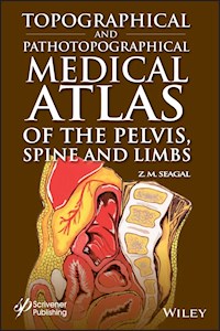
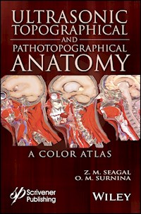
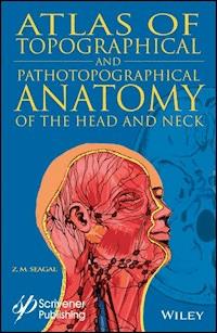
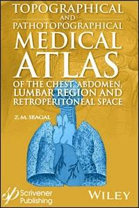


Table of Contents
Cover
Preface
Part 1: ULTRASONIC TOPOGRAPHICAL AND PATHOTOPOGRAPHICAL ANATOMY
1 Topography and Pathotopography of the Head
2 Topography and Pathotopography of the Neck
3 Topographical and Pathotopographical Anatomy of the Chest
4 Topographical and Pathotopographical Anatomy of the Abdomen
5 Topographical and Pathotopographical Anatomy of the Retroperitoneal Space
6 Topography and Pathotopography of the Pelvis
Topographical anatomy of the pelvic organs
Topography of the female pelvis
Topography of male pelvis
Ultrasonic topographical anatomy of male pelvis
7 Topography and Pathotopography of Lower Extremity
8 Conclusion
Part 2: TOPOGRAPHICAL AND PATHOTOPOGRAPHICAL MEDICAL ATLAS OF THE HEAD AND NECK
9 Introduction
10 The Head
Topographic Anatomy of the Head
Cerebral Cranium
Basis Cranii Interna
The Brain
Surgical Anatomy of Congenital Disorders
Pathotopography of the Cerebral Part of the Head
Facial Head Region
Dentes – Teeth
The Lymphatic System of the Head
Congenital Face Disorders
Pathotopography of Facial Part of the Head
11 The Neck
Topographic Anatomy of the Neck
Fasciae, Superficial and Deep Cellular Spaces and their Relationship with Spaces Adjacent Regions (Fig. 37)
Reflex Zones
Triangles of the Neck
Organs of the Neck (Fig. 50–51)
Pathography of the Neck
Topography of the neck
Part 3: TOPOGRAPHICAL AND PATHOTOPOGRAPHICAL MEDICAL ATLAS OF THE CHEST, ABDOMEN, LUMBAR REGION, AND RETROPERITONEAL SPACE
12 The Chest
Topographic Anatomy of the Chest
Chest Cavity Organs Projection and Layers of Chest
Surgical Anatomy of Thoracic Wall Congenital Malformation
Thoracic Cavity
Mediastinum Topography (Figures 17, 18, 19)
13 Abdomen
Topographic Anatomy of Anterolateral Abdomen Wall
Surgical Anatomy of Congenital Malformations of Anterior Lateral Abdominal Wall
Abdominal Region Topography
Abdominal Cavity Organs (Figures 45, 46)
14 Lumbar Region and Retroperitoneal Space
Topographic Anatomy of Lumbar Region and Retroperitoneal Space
15 PathotographyPathotographyof the Chest
Abdominal Cavity
Retroperitoneal Space
Part 4: TOPOGRAPHICAL AND PATHOTOPOGRAPHICAL MEDICAL ATLAS OF THE PELVIS, SPINE, AND LIMBS
16 Introduction
17 The Pelvis
Topographic Anatomy of the Pelvis
Individual, Gender and Age Differences:
The Organs of the Male Pelvis
The Topography of the Vas Deferens.
The Organs of the Female Pelvis
Defects of the Genitourinary System in Children
Perineum Topography
Pudendal Region in Men
Pudendal Region in Women
Surgical Anatomy of Congenital Pelvic and Perineum
Anus. Ischiorectal Fossa. Perineal Rectum
Surgical Anatomy of Congenital Malformations of the External Genitalia
Classification of Hypospadias
Pathotophography of the Peivis
18 The Spine
Topographic Anatomy of the Spine
Individual and Age Differences of the Spine
The Spinal Cord and Nerve Roots
Surgical Anatomy of the Malformations of the Spine and Spinal Cord
Pathotopography of the Spine
19 The Limbs
Topographic Anatomy of the Upper Limb
Shoulder
Forearm
Hand (Figure 81)
Surgical Anatomy of Congenital Malformations of the Upper Limb
Pathotopography of the Upper Limbs
Topographic Anatomy of Lower Limbs
Gluteal Region
Femur
Shin
Foot
Pathotopography of the Lower Limbs
Conclusion
Appendix A
Appendix B
End User License Agreement
List of Illustrations
Chapter 1
Figure 1 Layer-by-layer topography of the head.
Figure 2 Layer-by-layer topography.
Figure 3 Cross-section of the head.
Figure 4 Comparative ultrasonic topographical anatomy of the head.
Figure 5 Hollow depressed fracture.
Figure 6 Linear fracture.
Figure 7 Layer-by-layer ultrasonic topographical anatomy of the head.
Figure 8 Basilar region of the cranium.
Figure 9 Deep facial area.
Figure 10 Facial area of the head.
Figure 11 Infratemporal fossa.
Figure 12 Maxillary artery with medial meningeal artery and superficial temp...
Figure 13 Maxillary artery.
Figure 14 Mastication muscles.
Figure 15 Lymphatic system of the head and neck.
Figure 16 Linear fracture.
Figure 17 Tamponade of the ventricle of the brain IV with transition to pons...
Figure 18 Intraventricular blood clot.
Figure 19 Intraventricular hemorrhage.
Figure 20 Hematoma at the optic thalamus.
Figure 21 Frontobasal intracerebral hematoma.
Figure 22 Acute epidural hematoma of the parieto-occipital left region.
Chapter 2
Figure 23 Sagittal slice of the head and the neck.
Figure 24 Sagittal slice of the head and the neck.
Figure 25 Cross-section of the neck.
Figure 26 Muscles and fasciae of the neck.
Figure 27 Thyroid node.
Figure 28 Thyroid node (magnified).
Figure 29 Thyroid left lobe cysts.
Figure 30 Chronic thyroiditis.
Figure 31 Thyroid hemorrhagic cyst (magnified).
Figure 32 Common carotid artery sternosis.
Figure 33 Topography of carotid and vertebral arteries. Muscles and fasciae ...
Figure 34 Cyst with polyp.
Figure 35 Cyst with polyp (magnified).
Figure 36 Submaxillary lymphatic node.
Figure 37 Interfacial muscles of the neck.
Figure 38 Deep layers of the suprahyoid area.
Figure 39 Thyroid.
Figure 40 Topography of the scalenovertebral triangle.
Chapter 3
Figure 41 Sagittal section of the chest and abdomen.
Figure 42 Cross-section of the chest.
Figure 43 Topography of the mammary gland.
Figure 44 Topography of the chest.
Figure 45 Topography of the blood vessels and heart.
Figure 46 Topography of the blood vessels and heart.
Figure 47 Topography of the lung roots.
Figure 48 Topography of the bifurcation of the trachea and the aorta.
Figure 49 Topography of the heart and the lungs.
Figure 50 Topography of the heart.
Figure 51 Topography of the heart and blood.
Figure 52 Topography of the heart and blood vessels.
Figure 53 Dilatation of the cardiac chambers.
Figure 54 Mitral stenosis.
Chapter 4
Figure 55 Layer-by-layer topography of the anterior wall of the abdomen.
Figure 56 Frontal section of the abdominal cavity.
Figure 57 Cross-section at the level of the liver.
Figure 58 Topography of the abdomen.
Figure 59 Cross-section at the level of the liver.
Figure 60 Topography of the abdomen.
Figure 61 Frontal section at the level of chest and abdomen.
Figure 62 Topography of the anterolateral abdominal wall.
Figure 63 Syntopy of the abdominal cavity organs.
Figure 64 Arteries of the upper part of the abdominal cavity.
Figure 65 Arteries of the upper part of the abdominal cavity.
Figure 66 Arteries of the upper part of the abdominal cavity.
Figure 67 Liver.
Figure 68 Liver.
Figure 69 Liver.
Figure 70 Liver.
Figure 71 Liver.
Figure 72 Liver.
Figure 73 Gallbladder.
Figure 74 Spleen.
Figure 75 Spleen.
Figure 76 Spleen.
Figure 77 Spleen.
Figure 78 Lines of attachment of mesostenium and mesocolon.
Figure 79 Lines of attachment of mesostenium and mesocolon.
Figure 80 Liver pathology.
Figure 81 Tumor of the right hepatic lobe.
Figure 82 Liver concrement.
Figure 83 Hepatic cyst.
Figure 84 Gallbladder pathology.
Figure 85 Cholelithiasis.
Figure 86 Biliary system.
Figure 87 Gallbladder.
Figure 88 Abnormal development of the gallbladder.
Figure 89 Spleen pathology.
Figure 90 Abnormal development of the gallbladder.
Chapter 5
Figure 91 Retroperitoneal space organs.
Figure 92 Blood vessels of the retroperitoneal space.
Figure 93 Abdominal aorta and kidneys. Contrastless angiography.
Figure 94 Kidney.
Figure 95 Kidney.
Figure 96 Kidney.
Figure 97 Kidney.
Figure 98 Renal circulation.
Figure 99 Pancreas.
Figure 100 Chronic pancreatitis.
Figure 101 Pancreatic cyst.
Figure 102 Renal cyst.
Figure 103 Urolithiasis.
Figure 104 Renal tumor.
Figure 105 Renal hemangioma.
Chapter 6
Figure 106 Sagittal section of the pelvis.
Figure 107 Layer-by-layer frontal sections.
Figure 108 Layer-by-layer sections of blood vessels.
Figure 109 Frontal sections.
Figure 110 Cross-section of the pelvis.
Figure 111 Topography of the female pelvis.
Figure 112 Blood supply to internal female genital organs.
Figure 113 Sagittal cross-section of male pelvis.
Figure 114 Hysteromyoma.
Figure 115 Left ovarian cyst.
Figure 116 Ureteral opening calculus.
Figure 117 6 weeks pregnancy.
Figure 118 Calcification.
Figure 119 Left ureteral calculus.
Figure 120 Chronic prostatitis.
Chapter 7
Figure 121 Cross-section at the level of femur.
Figure 122 Cross-section at the level of shin bone.
Figure 123 Sagittal section at the level of foot.
Figure 124 Coxofemoral joint.
Figure 125 Sacroiliac joint.
Figure 126 Anterior superior iliac bone.
Figure 127 Femoral muscles.
Figure 128 Femoral bone.
Figure 129 Femoral head.
Figure 130 Femoral head.
Figure 131 Femoral artery. Femoral vein.
Figure 132 Suprapatellar pouch.
Figure 133 Infrapatellar recess.
Figure 134 Lateral recess.
Figure 135 Knee joint. Medial view.
Figure 136 Posterior recess.
Figure 137 Blood vessels of popliteal fossa.
Figure 138 Shin bone muscles, fibular bone.
Figure 139 Blood vessels of shin bone, posterior tibial muscle.
Figure 140 Shin bone, heel bone.
Figure 141 Ankle joint.
Figure 142 Foot.
Figure 143 Tarsal bones.
Figure 144 Main joint of toes.
Figure 145 Bones of great toe.
Figure 146 Aseptic necrosis of femoral head.
Figure 147 Rheumatoid arthritis.
Figure 148 Rheumatoid arthritis.
Figure 149 Rheumatoid arthritis.
Figure 150 Rheumatoid arthritis.
Figure 151 Rheumatoid arthritis.
Figure 152 Rheumatoid arthritis.
Figure 153 Rheumatoid arthritis.
Chapter 10
Figure 1 Vessels and nerves of the head.
Figure 2 Layers of the frontal-parietal-occipital area.
Figure 3 Regio temporalis.
Figure 4 Mastoid process with Shipo’s triangle.
Figure 5 Inner cranium base.
Figure 6 Medial section of head and neck
Figure 7 Branches of trigeminal nerve
Figure 8 Scheme of head skin innervation.
Figure 9 Superficial head formations.
Figure 10 Branches of the facial nerve
Figure 11 Cerebral vessels. MRI.
Figure 12 Cerebral vessels
Figure 13 Dural venous sinuses
Figure 14 Dural venous sinuses. MRI.
Figure 15 Dural venous sinuses.
Figure 16 Venous sinuses of the dura mater.
Figure 17 Bleeding in the brain due to rupture of the aneurism
Figure 18 Types of intracranial hemorrhage
Figure 19 Different types of brain hematomas
Figure 20 Orbital muscles, vessels and nerves. Topdown view,
Figure 21 Orbital muscles, vessels and nerves. Side view.
Figure 22 The nasal cavity
Figure 23 Variants of pneumatization of maxillar sinuses depending of maxill...
Figure 24 Buccal region.
Figure 25 Profound face area.
Figure 26 Profound face area.
Figure 27 Dural arteries. Side view
Figure 28 Cellular spaces of profound face areas
Figure 29 Regi o infraorbitalis
Figure 30 Regio infraorbitalis
Figure 31 Infraorbital region
Figure 32 Localization of phlegmons
Figure 33 Purulent parotitis
Figure 34 Pannus formation
Figure 35 Pannus of nasopharynx
Chapter 11
Figure 36 Neck area
Figure 37 Fascias of neck
Figure 38 The lymphatic system of the head and neck
Figure 39 Areas of triangles and neck
Figure 40 Deep layers suprahyoid area
Figure 41 Side Triangle neck
Figure 42 External carotid artery
Figure 43 Options relations bifurcation of the common carotid arteries and v...
Figure 44 Innervation and blood supply of the neck muscles
Figure 45 Surface vessels and nerves of the neck
Figure 46 The branches of the cervical plexus
Figure 47 Surface education neck
Figure 48 Deep neck Education
Figure 49 The topography of organs, blood vessels and nerves of the neck
Figure 50 Topography staircase-vertebral triangle
Figure 51
Figure 52 The cut at the level of the thyroid isthmusNewborn:
Figure 53 Typical localization of abscesses and abscesses of the neck
Figure 54 Patotopographic anatomy of the neck
Figure 55 Ectopic and normal arrangement of the various types of goiter
Figure 56 Nodular goiter of the thyroid gland
Figure 57 Thyroid nodular goiter with compression of the larynx and trachea...
Chapter 12
Figure 1 Transverse section of the chest. Diaphragm.
Figure 2 Lung segments.Lobus superior:
Figure 3 Thymus gland and its connection with the thyroid gland.
Figure 4 Differences in the shape and number of thymus glands.
Figure 5 Topography of blood vessels, heart, right lung.
Figure 6 The relationship of the trachea, esophagus and aorta. Topography of...
Figure 7 Layers of the front upper region of the sternum.
Figure 8 Vessels and nerves of the chest wall.
Figure 9 Vessels and nerves of the chest wall.
Figure 10 Intercostal space.
Figure 11 Mammary gland.
Figure 12 MRI and CT of the breast.
Figure 13 Diaphragm.
Figure 14 Diaphragm. Vessels.
Figure 15 Right lung.
Figure 16 Left lung.
Figure 17 Posterior mediastinum, left view.
Figure 18 Posterior mediastinum, right view.
Figure 19 Transverse section of the breast.
Figure 20 Organs of anterior mediastinum and neck.
Figure 21 Syntopia of the chest cavityorgans and neck.
Figure 22 Infant’s pericardium.
Figure 23 Topography of blood vessels, heart, right lung.
Figure 24 Heart and lungs.
Figure 25 Heart.
Figure 26 Heart. Back view.
Figure 27 Syntopia of heart, big vessels and lungs.
Figure 28 The relationship of the trachea, esophagus and aorta.
Figure 29 Pattern of segmental division of the bronchial tree.
Figure 30 Posterior mediastinum.
Figure 31 Blood supply of the middle and lower parts of the chest cavity.
Figure 32 Types of esophagus atresia.
Figure 33 Topography of the organs of the posterior mediastinum. Front view....
Figure 34 Organs and lymph nodes of the mediastinum.
Chapter 13
Figure 35 Sections of anterior lateral abdomen wall.
Figure 36 Transversal sections of anterior abdominal wall at different level...
Figure 38 Topography of inferior section of anterior abdominal wall.
Figure 39 Anterior abdominal wall blood supply and anastomoses.
Figure 40 Internal surface of anterior abdominal wall. Removed peritoneum in...
Figure 41 The topography of the anterolateral abdominal wall.
Figure 42 Content of hepatoduodenal copula.
Figure 43 The organs of the abdominal cavity. The upper floor of the abdomin...
Figure 44 The ground floor of the abdominal cavity.
Figure 45 Syntopy of abdominal organs.
Figure 46 Holotopy of abdominal organs in newborns.
Figure 47 The projection of the liver and stomach to the anterior abdominal ...
Figure 48 Arteries of the upper abdomen.
Figure 49 Vessels submucosal plexus of the fundus (transillumination angiogr...
Figure 50 Anastomoses between intramural vessels submucosal plexus of the an...
Figure 51 The serous-muscular layer of the stomach of the dog in transmitted...
Figure 52 The digested stomach reflected light (experiment on dogs).
Figure 53 The topography of the duodenum.
Figure 54 Syntopy of the duodenum and pancreas.
Figure 55 Organs of the abdominal cavity. Anterior abdominal wall of the sto...
Figure 56 The lines of attachment mesentericof thin intestine and colon.
Figure 57 Various forms of the cecum and the appendix in the newborn.
Figure 58 Blood supply of the colon.
Figure 59 Blood supply of the sigmoid colon and rectum.
Figure 60 Features of the blood supply of the large intestine in the newborn...
Figure 61 Liver.
Figure 62 Visceral surface of the liver and its topography.
Figure 63 Syntopy of liver and colon. The front wall of the stomach, small i...
Figure 64 The topography of the nerves of the diaphragm.
Figure 65 Diaphragma.
Figure 66 Liver of the newborn. The dashed line indicates the edge of the co...
Figure 67 Non contrastive transillumination angiogram gallbladder.
Figure 68 Transillumination pigment angiogram distal gallbladder.
Figure 69 Gallbladder newborn.
Figure 70 The scheme of the biliary tract.
Figure 71 Differences of vesico-colonic ligament.
Figure 72 Pancreas.
Chapter 14
Figure 73 Lumbar region.
Figure 74 Topography of retroperitoneal space.
Figure 75 The posterior wall of the abdomen.
Figure 76 The organs and major blood vessels of the retroperitoneal space....
Figure 77 Structure of the kidney.
Figure 78 The blood vessels of kidneys and adrenal glands.
Figure 79 Kidney Vessels.
Figure 80 Kidney topography.
Figure 81 Entrance to the small pelvis of the newborn girl, front and top vi...
Chapter 15
Figure 82 Mammary gland.
Figure 83 Mammary gland.
Figure 84 Transverse section of the chest.
Figure 85 Mediastinum after removal of the right lung. Right view.
Figure 86 Transverse section of the chest.
Figure 87 Cross section of the thorax at the level of the liver. 1,3,5 – lob...
Figure 88 Cross section of the thorax at the level of the liver.
Figure 89 Hemangioma of the liver.
Figure 90 Tumor of the right lobe of the liver.
Figure 91 Additional lobe of the spleen.
Figure 92 Anomaly of development of the gallbladder.
Figure 93 Cataral cholecystitis (transillumination angiograms).
Figure 94 Destructive forms of cholecystitis.
Figure 95 Planar shadow image of a cancerous focus.
Figure 96 Saucer-like stomach cancer.
Figure 97 Zones of possible growth and spread of tumor.
Figure 98 Cancer of the cardiac part of the stomach with passage to the esop...
Figure 99 Polyposis of the large intestine. Transillumination angiograms in ...
Figure 100 Chronic ulcerative colitis.
Figure 101 Sagittal male pelvis.
Figure 102 Cyst of pancreas.
Figure 103 Cyst of pancreas.
Figure 104 Chronic pancreatitis.
Figure 105 Chronic pancreatitis.
Figure 106 Renal neoplasms.
Figure 107 Tumor of the kidney.
Figure 108 Hemangioma of the kidney.
Figure 109 Urolithiasis disease.
Chapter 17
Figure 1 Urogenital diaphragm in women.
Figure 2 The front section of the pelvis.
Figure 3 The innervation of the hip joint.
Figure 4 The innervation of the hip joint.
Figure 5 Urogenital diaphragm in men.
Figure 6 The Pelvic Plexus.
Figure 7 Topography of the bladder.
Figure 8 Sagittal section of the male pelvis.
Figure 9 Cross section of the male pelvis.
Figure 10 The bladder and the prostate.
Figure 11 Median sagittal section of the pelvis of a newborn boy.
Figure 12 Arteries and veins of the male pelvis.
Figure 13 The prostate and the seminal vesicles.
Figure 14 Transillumination angiogram of the broad ligament.
Figure 15 Blood supply of female reproductive organs.
Figure 16 Arteries and veins of the female pelvis.
Figure 17 The topography of the female pelvis. The uterus and vagina at the ...
Figure 18 Sagittal section of the female pelvic organs.
Figure 19 The round ligament of the uterus in transmitted light.
Figure 20 Topography of the male perineum.
Figure 21 Topography of the female perineum.
Figure 22 Median sagittal section of the newborn girl pelvis. A right side v...
Figure 23 Shell of testis and layers of the scrotum.
Figure 24 Topography of muscles and fascia perineum in a newborn girl.
Figure 25 Sagittal section of pelvic girl 5 months old.
Figure 26 Sagittal section of pelvic girls 2 years old.
Figure 27 Pelvic organs of newborn girl.
Figure 28 Rectal atresia.
Figure 29 Sagittal section of pelvic of a boy, 5 years.
Figure 30 Pelvis organs if newborn boy.
Figure 31 Topography of muscles and fascia of the perineum of newborn boy....
Figure 32 Uterine fibroids.
Figure 33 Cyst of the left ovary.
Figure 34 Cross section of a female small pelvis.
Figure 35 Sagittal cutting female pelvis.
Figure 36 Cross section of the female pelvic organs.
Figure 37 Sagittal section of the female pelvic organs. MRI.
Figure 38 Interrelation of the organs of the female pelvis.
Figure 39 Sagittal section of the male pelvic organs.
Figure 40 Sagittal cut male pelvis.
Figure 41 Cross section of the male pelvic organs.
Figure 42 Cross section of the male pelvis.
Figure 43 Localization of ulcers in the cellular spaces of the pelvis: anter...
Chapter 18
Figure 44 Sagittal section of the chest.
Figure 45 Sagittal section of the female pelvic organs.
Figure 46 Sagittal section of the cervical processes of the vertebrae.
Figure 47 Pelvic plexus.
Figure 48 The sciatic nerve and its branches.
Figure 49 Vessels of the brain. MRI.
Figure 50 Sagittal section of the head and neck.
Figure 51 Typical localization of abscesses and phlegmon of the neck.
Figure 52 MRI. MRI of the cervical spine (hernia).
Figure 53 Neck pathography.
Figure 54 Sagittal section of the chest.
Figure 55 Sagittal section of the chest.
Chapter 19
Figure 56 The brachial plexus.
Figure 57 The axillary area.
Figure 58 Lymphatic vessels of axilla.
Figure 59 Deltoid region.
Figure 60 Different forms of trigonum clavipectorale (by D.D. Kupatadze).
Figure 61 Blood circulation of the scapular region.
Figure 62 Crosscut of shoulder in the lower third.
Figure 63 Crosscut of shoulder in the lower third (MRI, CT).
Figure 64 The posterior brachium area.
Figure 65 The topography of the anterior brachium area.
Figure 66 The topography of the radial nerve and the deep artery of the uppe...
Figure 67 The front surface of the forearm.
Figure 68 Anterior ulnar area.
Figure 69 The posterior ulnar area.
Figure 70 The posterior area of the forearm.
Figure 71 Muscles of the upper limb. Front view.
Figure 72 Arteries of the upper limb.
Figure 73 Crosscut of the left forearm in the middle third.
Figure 74 The front surface of the left forearm.
Figure 75 The front surface of the right forearm.
Figure 76 Topography of the upper third of the right forearm.
Figure 77 Subcutaneous nerves of the upper limb. Front view.
Figure 78 Subcutaneous nerves of the upper limb. Back view.
Figure 79 Subcutaneous lymphatic and venous vessels of the upper limb.
Figure 80 Palm surface of the wrist joint.
Figure 81 Frontal section of the hand.
Figure 82 The topography of the surface palm formations.
Figure 83 The deep palmar structures.
Figure 84 Posterior surface of the pulm.
Figure 85 Subcutaneous structures of the palm.
Figure 86 Topography of the palmar surface of the wrist joint.
Figure 87 The topography of the veins on the palmar surface of the wrist joi...
Figure 88 Topography of the palm.
Figure 89 Fingers.
Figure 90 Cross section of the shoulder. Osteomyelitis of humerus.
Figure 91 Cross section of the shoulder. Osteomyelitis of the humerus.
Figure 92 Pathotopographic anatomy of the transverse section of the shoulder...
Figure 93 Frontal section of the hand.
Figure 94 Gluteal region.
Figure 95 The sciatic nerve and its branches. Back view.
Figure 96 Blood supply and innervation of the gluteal muscles.
Figure 97 Blood supply and innervation of the gluteal muscles.
Figure 98 The topography of the gluteal region.
Figure 99 Muscles of the lower limb. Front view.
Figure 100 The femoral triangle.
Figure 101 Blood supply and innervation of the back of the thigh.
Figure 102 The posterior thigh area.
Figure 103 Muscles of the lower limb. Back view.
Figure 104 Channel adductors.
Figure 105 The cross-cut of the thigh.
Figure 106 Cross-cut thigh (MRI and CT).
Figure 107 Vascular and neuromuscular lacunas.
Figure 108 The femoral canal.
Figure 109 The lower extremity arteries. Front view.
Figure 110 Topography of the popliteal fossa.
Figure 111 Cross section of the thigh. The cartilaginous tumor, cancer «
in s
...
Figure 112 Cross section of the thigh. The cartilaginous tumor, cancer «
in s
...
Figure 113 Cross section of the thigh. Mesenchymal fibrosarcoma of thigh.
Figure 114 Cross section of the thigh. Mesenchymal fibrosarcoma of thigh. (M...
Figure 115 The relationship of the femoral head and the acetabulum of the hi...
Figure 116 The front surface of the shin.
Figure 117 The posterior area of the shin.
Figure 118 The cross-cut of the upper third of the shin.
Figure 119 Topography of the upper third of the back of the shin.
Figure 120 Topography - vascular nerve bundle upper third of the back of the...
Figure 121 A cross section of the shin.
Figure 122 A cross section of the shin (CT).
Figure 123 The knee joint. Front view.
Figure 124 The knee joint. The view from the medial side.
Figure 125 The knee joint. View from the lateral side.
Figure 126 Volvulus of the right knee.
Figure 127 Sagittal section of the foot.
Figure 128 Sagittal section of the foot.
Figure 129 Sagittal section of the foot (MRI).
Figure 130 Area of the inner ankle.
Figure 131 The sole of the foot.
Figure 132 Surface vessels and nerves of the ankle and posterior foot.
Figure 133 Anterior area of the ankle joint.
Figure 134 Topography of heel.
Figure 135 Topography of the medial surface of the ankle joint.
Figure 136 Subcutaneous veins and nerves of the lower extremities.
Figure 137 Superficial lymph vessels of the lower limb. Back view.
Figure 138 Superficial lymph vessels of the lower limb. Front view.
Figure 139 Transverse section of the thigh. Mesenchymal fibrosarcoma of the ...
Figure 140 Pathotopography of the transverse section of the thigh in the MR-...
Figure 141 Transverse section of the thigh. Cartilage tumor, cancer “
in situ
Figure 142 Transverse section of the femur on CT. Cartilage tumor, cancer “
i
...
Figure 143 Sagittal section of the foot on the MR-tomogram.
Figure 144 Sagittal section of foot.
Guide
Cover
Table of Contents
Begin Reading
Pages
ii
iii
iv
ix
x
xi
1
3
4
5
6
7
8
9
10
11
12
13
14
15
16
17
18
19
20
21
22
23
25
26
27
28
29
30
31
32
33
34
35
36
37
38
39
40
41
43
44
45
46
47
48
49
50
51
52
53
54
55
56
57
58
59
60
61
62
63
64
65
66
67
68
69
70
71
72
73
74
75
76
77
78
79
80
81
82
83
84
85
86
87
88
89
91
92
93
94
95
96
97
98
99
100
101
102
103
104
105
106
107
108
109
110
111
112
113
114
115
116
117
118
119
120
121
123
124
125
126
127
128
129
130
131
132
133
134
135
136
137
138
139
140
141
142
143
144
145
146
147
148
149
150
151
152
153
154
155
156
157
159
160
161
163
164
165
166
167
168
169
170
171
172
173
174
175
176
177
178
179
180
181
182
183
184
185
186
188
187
189
190
191
192
193
194
195
196
197
198
199
200
201
202
203
204
205
206
207
208
209
210
211
212
213
214
215
216
217
218
219
220
221
222
223
224
225
226
227
228
229
230
231
232
233
234
235
236
237
238
239
240
241
242
243
244
245
246
247
248
249
250
251
252
253
254
255
256
257
258
259
260
261
262
263
264
265
267
268
269
270
271
272
273
274
275
276
277
278
279
280
281
282
283
284
285
286
287
288
289
290
291
292
293
294
295
296
297
298
299
300
301
302
303
304
305
306
307
308
309
310
311
312
313
314
315
316
317
319
321
322
323
324
325
326
327
328
329
330
331
332
333
334
335
336
337
338
339
340
341
342
343
344
345
346
347
348
349
350
351
352
353
354
355
356
357
358
359
360
361
362
363
364
365
366
367
368
369
371
372
373
374
375
376
377
378
379
380
381
382
383
384
385
386
387
388
389
390
391
392
393
394
395
396
397
398
399
400
401
402
403
404
405
406
407
408
409
410
411
412
413
414
415
416
417
418
419
420
421
422
423
424
425
426
427
428
429
430
431
432
433
434
435
436
437
438
439
440
441
442
443
444
445
446
447
448
449
450
451
452
453
454
455
456
457
458
459
460
461
462
463
464
465
466
467
468
469
470
471
472
473
474
475
476
477
478
479
480
481
482
483
484
485
486
487
488
489
490
491
492
493
494
495
496
497
498
499
501
502
503
504
505
506
507
508
509
510
511
512
513
514
515
516
517
518
519
520
521
522
523
524
525
526
527
528
529
530
531
532
533
534
535
536
537
538
539
540
541
542
543
544
545
546
547
548
549
550
551
552
553
554
555
556
557
558
559
560
561
562
563
564
565
566
567
568
569
570
571
572
573
574
575
576
577
578
579
580
581
582
583
584
585
586
587
588
589
590
591
593
594
595
596
597
598
599
600
601
602
603
604
605
606
607
608
609
610
611
612
613
614
615
616
617
618
619
620
621
622
623
624
625
626
627
628
629
630
631
632
633
634
635
636
637
638
639
640
641
642
643
644
645
646
647
648
649
650
651
652
653
654
655
656
657
658
659
660
661
662
663
664
665
666
667
668
669
670
671
672
673
674
675
676
677
678
679
680
681
682
683
684
685
687
688
689
690
691
692
693
694
695
696
697
698
699
700
701
702
703
704
705
706
707
710
711
712
713
714
715
716
Scrivener Publishing100 Cummings Center, Suite 541JBeverly, MA 01915-6106
Publishers at ScrivenerMartin Scrivener ([email protected])Phillip Carmical ([email protected])
Topographical and Pathotopographical Medical Atlas of the Human Body
Z. M. Seagal
This edition first published 2020 by John Wiley & Sons, Inc., 111 River Street, Hoboken, NJ 07030, USA and Scrivener Publishing LLC, 100 Cummings Center, Suite 541J, Beverly, MA 01915, USA© 2020 Scrivener Publishing LLCFor more information about Scrivener publications please visit www.scrivenerpublishing.com.
All rights reserved. No part of this publication may be reproduced, stored in a retrieval system, or transmitted, in any form or by any means, electronic, mechanical, photocopying, recording, or otherwise, except as permitted by law. Advice on how to obtain permission to reuse material from this title is available at http://www.wiley.com/go/permissions.
Wiley Global Headquarters111 River Street, Hoboken, NJ 07030, USA
For details of our global editorial offices, customer services, and more information about Wiley products visit us at www.wiley.com.
Limit of Liability/Disclaimer of WarrantyWhile the publisher and authors have used their best efforts in preparing this work, they make no representations or warranties with respect to the accuracy or completeness of the contents of this work and specifically disclaim all warranties, including without limitation any implied warranties of merchant-ability or fitness for a particular purpose. No warranty may be created or extended by sales representatives, written sales materials, or promotional statements for this work. The fact that an organization, website, or product is referred to in this work as a citation and/or potential source of further information does not mean that the publisher and authors endorse the information or services the organization, website, or product may provide or recommendations it may make. This work is sold with the understanding that the publisher is not engaged in rendering professional services. The advice and strategies contained herein may not be suitable for your situation. You should consult with a specialist where appropriate. Neither the publisher nor authors shall be liable for any loss of profit or any other commercial damages, including but not limited to special, incidental, consequential, or other damages. Further, readers should be aware that websites listed in this work may have changed or disappeared between when this work was written and when it is read.
Library of Congress Cataloging-in-Publication Data
ISBN 978-1-119-61433-3
Cover image: 3D Illustration, Pepe Gallardo | Dreamstime.com & Abstract Background, Pokaz | Dreamstime.comCover design by Kris Hackerott
Preface
Topographical and Pathotopographical Medical Atlas of the Human Body is a fundamental and practically important book designed for doctors of all specializations and students at medical schools. The atlas contains almost everything connected with the topographic and pathotopographic human anatomy, topography of different areas in layers, pathotopography, variant, normal, computer and magnetic resonance imaging (MRI) of topographic and pathotopographic anatomy. Also presented in the atlas are new theoretical and practical sections of topographic anatomy developed by the author; these are published for the first time. They are of practical importance in mastering the technique of operative interventions and denying the possibility of iatrogenic complications during operations.
Images of individual and age-related changes in human organs are presented according to variant topographic anatomy. Topographic anatomy describes holotopy, skeletopy, organ syntopy and main anatomic formations, fascial compartments, cellular spaces, neurovascular bundles, collateral circulation, sensor and motor parts of nerve trunks. Pathotopographical anatomy is shown on typical pathology examples in various fields.
It is important to visualize topographical anatomy structures not only on a corpse, but also on patients who have undergone surgical interventions. These data were obtained using computerized MRI topography, transillumination of organs and tissues, pathotopography and ultrasound topography.
Currently, there are no modern atlases in the world for clinical topographic and pathotopographic anatomy. There are several reasons for this. Firstly, this subject has been studied only in Russia and Hungary. In other countries, it is either absent or present in related disciplines, such as anatomy or surgery. A number of sections, such as pathotopographic, variant, normal topographic anatomy, transillumination, and vital topographic and pathotopographic anatomy on sick and healthy people were developed by the author of this atlas for the first time.
This atlas can serve as reference in the daily work of theoretical and practical physiologists and pathophysiologists, anatomists and typographic anatomists, pathologists, therapists, surgeons, obstetricians and gynecologists, neurologists and dermatovenerologists, radiation diagnostitions, anesthesiologists, otolaryngologists, trauma surgeons, ortho-pedologists, pediatricians, and dentists, as well as those in narrow specialties, such as cardiovascular surgeons, urologists, plastic surgeons, neurosurgeons, etc.
The topographic and pathotopographic anatomy presented can be subdivided into surgical, anatomy, therapy, dental and pediatric. Specific and non-specific features of pathotopography and normal variants have already been found and are presented in the atlas; normal anatomy, divided into pathological, ultrasound topographic and pathotopographic anatomy, is established in the book.
Topographic clinical anatomy changes with the disease and recovery during iatrogenic manipulations, surgical and therapeutic treatment. That is why some of these points are described in the atlas.
Our atlas of topographic and pathotopographic anatomy is a full-colored handbook for students and doctors in almost all specialties. It covers all the organs and tissues of the human body. Its fundamental and practical importance is that topography and patho-topograpy are presented for the first time in vivo. These sections (ultrasound topography and pathotopography, transillumination topography and pathotopography) are of great practical importance both for early diagnosis and for providing the right treatment, including proper surgical tactics. They are important for surgeons in planning and performing numerous surgical operations. The author of the atlas has a unique museum of operative surgery at the Academy, which is visited by doctors and students from all over the world. Some exhibits from this museum are presented in our atlas. They are normal and pathotopographical surgical anatomy, electrified stands and steps of surgical intervention on the complex of formalin preparations. This three-track museum is dedicated to typical and atypical surgical interventions, as well as the author´s operations with use of the author´s original tools and techniques. Nothing like this currently exists in the world. It is reflected in the atlas and has great educational and practical importance both in surgery and medicine. There are more than a hundred complexes in the museum which were developed and collected on the operations on various organs and tissues in surgery, dentistry, pediatric surgery, thoracic surgery, urology, and other areas and organs. Thus, surgical anatomy became particular for practical doctors of various specialties. In modern medicine, major problems are late diagnosis, false positive and false negative diagnostic results, and stage and size of acute pathology, which can lead to wrong treatment, mortality and disability of patients, and also to recurrence of the disease, ineffective treatment, wrong prognosis and incorrect prevention of post-operative complications in the short term and the long term. A gold standard of diagnostic and treatment, unfortunately, does not include appropriate and effective control of the diagnosis and treatment, which needs to be developed.
Comparative analysis of СT scan, MRI and pathotogram, including vital transilluminatory and ultrasound ones, revealed benefits of our vital monitoring method, which is reflected in the atlas. It provides more information and specifics on malignant and benign tumor, on degenerative, dystrophic, inflammatory processes, traumas and its complications. In the atlas there are clinical data reversible and irreversible of regional blood circulation disorder with focal pathology (adenoma, cyst, breast and thyroid cancer, vessel stenosis, intestinal paresis, etc.). Methods like PET, MRI and X-ray are highly sensitive but they cannot guide a doctor live during an operation in tumor topography. Maintenance of the surgery with these methods is not possible, which leads to iatrogenic complications: bleed, damage of lactiferous duct, and false results, and also causes an increase in the extent of the operation, recurrence and other complications. Pathotopograms mentioned in the atlas are important to determine surgical access, to visualize normal and changed lymph nodes, and to know the required extent of the operation. Ultrasound pathotopograms of focal arthritis and rheumatoid arthritis mentioned in the atlas revealed new specific differential signs of pathology, which is of practical importance in the treatment and prevention of iatrogenic rheumatic complications and in screening studies.
Our work is devoted to the memory of patients who have died from the lack of scientific achievements, lifetime pathotopograms and a new diagnostic technology. One might be surprised at the very possibility of such support in the pre-operative diagnosis, surgery and postoperative treatment. But this technological support not only accompanies, but also helps the surgeon to choose between conservative and other tactics of treating the patient. We hope that this full-color book will prolong life in patients and improve the skill of doctors and students in diagnosing and preventing diseases. Between the doctor and the patient should stand medical equipment, but it should not block them from each other. A doctor armed only with a scalpel is dangerous for the patient. A doctor armed with modern and original technology is dangerous for his illness.
Managers of our pathotopographic, transilluminative and ultrasound technology already operate in urgent and scheduled medicine. Our atlas and devices will be used by future doctors. Now is the time to speed up and implement rational innovations, and so it is necessary to start with the government, rather than finish with it. Three periods of illness – preoperative, surgery and post-operative, as well as three periods of life – childhood, maturity and old age are inseparable from each other, because they are associated with one person, one illness. As in life a wise mentor is needed during treatment, surgery and the post-operative period; this cannot be accomplished without the wise, lifetime functional pathotopographic results presented in our atlas. Flexible new medical technologies presented to the reader put them in a position to guarantee the patient’s vitality.
Part 1ULTRASONIC TOPOGRAPHICAL AND PATHOTOPOGRAPHICAL ANATOMY
1Topography and Pathotopography of the Head
The chapter on the ultrasonic topography and pathotopographical anatomy of the head includes layer-by-layer topography of the visceral and cerebral craniums with the cross-sectional imaging of the head.
Ultrasonic images of external and internal bone lamellae, vessels of the subcutaneous layer, skin, and subcutaneous fat, depressed compression and linear fractures are demonstrated. Ultrasonic images of the medial cerebral artery, infundibulum, posterior communicating artery, pons cerebelli, medulla oblongata, anterior inferior cerebellar artery, basilar artery, anterior cerebral artery, posterior cerebral artery, and olfactory tract are verified based on the topographical anatomy of the basilar region of the cranium.
The deep facial area contains the internal wing muscle, mandibulum, and submandibular salivary gland; the oral cavity contains the tongue, peripharyngeal space, and posterior veil of the soft palate, as well as the superficial temporal artery, auriculotemporal nerve, maxillary artery, and middle meningeal artery. The ultrasonic images of the internal and external muscles are shown.
Images of the parotid gland, superficial cervical lymph nodes, and common carotid artery are presented.
Linear fracture is associated with the external bone lamella of the area of intact bone, with the intracranial space, and the hypoechogenic track. Under conditions of tamponade of the fourth ventricle of cerebrum with transition to the pons cerebelli, a blood clot is revealed in the vicinity of the clinoid plate at the pyramid apex of the temporal bone. The intraventricular blood clot can be pathotopographically associated with the left lateral ventricle, whereas liquid blood is observed at the lumen of the right lateral ventricle.
The atlas also contains images of the pathotopographical anatomy of the intraventricular hemorrhage, hematoma in the thalamus, frontobasal intracerebral hematoma, and acute epidural hematoma in the left parieto-occipital space accompanied by the phenomenon of the “boundary amplification”.
Thus, the ultrasonic topographic anatomy of the head provides the basis for the research into the pathotopographical anatomy of a given pathology and determines specific diagnostic features of injuries and/ or volume structures.
Figure 1 Layer-by-layer topography of the head.
1. v. brachiocephalica; 2. v. jugularis anterior sinistra; 3. v. jugularis anterior dextra; 4. v. angularis; 5. Plexus cervicalis; 6. v. supratrochlearis; 7. v. nasofrontalis; 8. a. angularis; 9. v. temporalis superlicialis; 10. n. supraorbitalis; 11. n. auriculotemporalis; 12. Plexus cervixalis; 13. m. depressor labii inferioris; 14. m. dieastricus (venter anterior).
Figure 2 Layer-by-layer topography.
1. Falx cerebri; 2. Sinus maxillaries; 3. n. hypoglossus; 4. n. hypoglossus; 5. n. facialis; 6. n. lingualis; 7. n. alveolaris inferior; 8. Arcus maxillae inferioris; 9. m. temporalis
Figure 3 Cross-section of the head.
1. Eye bulb; 2. Optic nerve; 3. Temporal lobe; 4. Internal carotid artery; 6. Pons varolii; 7. Ventricle of the brain IV; 8. Cerebellar hemisphere; 9. Optic nerve; 10. Visual tract; 11. Middle cranial fossa; 12. Temporal lobe of cerebral hemisphere; 13. Ephippium; 16. Temporal gyrus.
Figure 4 Comparative ultrasonic topographical anatomy of the head.
1. Subcutaneous tissue; 2. Vessels of the subcutaneous layer; 3. Skin; 5. Subgaleal cellular tissue; 8. Bone.
Figure 5 Hollow depressed fracture.
Figure 6 Linear fracture.
Figure 7 Layer-by-layer ultrasonic topographical anatomy of the head.
1. Subcutaneous tissue; 2. Vessels of the subcutaneous layer; 3. Skin; 4. Tendinous intersections; 5. Subgaleal cellular tissue; 6. Aponeurosis; 7. Subperiosteal cellular tissue; 8. Bone; 9. Pacchionian granulations; 10. Epidural cavity; 11. Subdural space; 12. Arachnoid membrane; 13. Choroid; 14. Venous sinus; 15. Encephalon; 16. Subarachnoid space; 17. Dura mater; 18. Periosteal coverage; 19. Diploic vein and draining vein.
Figure 8 Basilar region of the cranium.
1. Medial cerebral artery; 2. Infundibulum; 3. Posterior communicating artery; 4. Cerebral peduncles; 5. Pons cerebelli; 6. Vertebral artery; 7. Medulla oblongata; 8. Occipital sinus; 9. Posterior inferior artery of cerebellum; 10. Anterior inferior artery of cerebellum; 11. Basilar artery; 12. Posterior cerebral artery; 13. Interior carotid artery; 14. Optic chiasma; 15. Anterior communicating artery; 16. Anterior cerebral artery; 17. Olfactory tract.
Figure 9 Deep facial area.
1. Temporal muscle; 2. Temporal pterygoid space; 3. Buccal nerve; 4. Arcus zygomaticus; 5. Exterior pterygoid muscle; 6. Masticatory muscle; 7. Interior pterygoid muscle; 8. Mandible; 9. Subglossal cellular space; 10. Bed of submandibular salivary gland; 11. Submandibular salivary gland; 12. Mylohyoid muscle; 13. Tongue; 14. Peripharyngeal space; 15. Soft palate; 16. Masticator maxillary space.
Figure 10 Facial area of the head.
1. Temporal muscle; 2. Temporal pterygoid space; 3. Buccal nerve; 4. Arcus zygomaticus; 5. Exterior pterygoid muscle; 6. Masticatory muscle; 7. Interior pterygoid muscle; 8. Mandible; 9. Subglossal cellular space; 10. Bed of submandibular salivary gland; 11. Submandibular salivary gland; 12. Mylohyoid muscle; 13. Tongue; 14. Peripharyngeal space; 15. Soft palate; 16. Masticator maxillary space.
Figure 11 Infratemporal fossa.
1. Temporal muscle; 2. Temporal pterygoid space; 3. Buccal nerve; 4. Arcus zygomaticus; 5. Exterior pterygoid muscle; 6. Masticatory muscle; 7. Interior pterygoid muscle; 8. Mandible; 9. Subglossal cellular space; 10. Bed of submandibular salivary gland; 11. Submandibular salivary gland; 12. Mylohyoid muscle; 13. Tongue; 14. Peripharyngeal space; 15. Soft palate; 16. Masticator maxillary space.
Figure 12 Maxillary artery with medial meningeal artery and superficial temporal artery.
1. Superficial temporal artery and auriculotemporal nerve; 2. Internal maxillary artery; 3. Facial nerve; 4. Medial meningeal artery; 5. Exterior carotid artery; 6. Inferior alveolar nerve; 7. Lingual nerve; 8. Submental artery and nerve; 9. Buccal nerve; 10. Suborbital artery.
Figure 13 Maxillary artery.
1. Superficial temporal artery and auriculotemporal nerve; 2. Maxillary artery; 3. Facial nerve; 4. Medial meningeal artery; 5. Exterior carotid artery; 6. Inferior alveolar nerve; 7. Lingual nerve; 8. Submental artery and nerve; 9. Buccal nerve; 10. Suborbital artery.
Figure 14 Mastication muscles.
1. M. masseter; 2. A. masseterica; 3. A. carotis externa; 4. Mandibular dipole; 5. N. auriculotemporalis; 6. Collum mandibulae; 7. M. temporalis; 8. A. et n. temporalis profunda; 9. A. buccalis; 10. M. pterigoideus externus; 11. Ramus marginalis mandibulae; 12. M. pterigoideus internus.
Figure 15 Lymphatic system of the head and neck.
1. Parotid gland; 2. Occipital lymph nodes; 3. Posterior auricular lymph nodes; 4. Deep superior cervical glands; 5. Internal jugular; 6. Superficial cervical glands; 7. Deep inferior cervical glands; 8. Anterior auricular lymph nodes; 9. Mandibular lymph nodes; 10. Submental lymph nodes; 11. Common carotid artery; 12. Right jugular lymph trunk.
Figure 16 Linear fracture.
1. External bone lamella at the region of intact bone; 2. Intracranial space; 3. Linear fracture; 4. Hypoechogenic track.
Figure 17 Tamponade of the ventricle of the brain IV with transition to pons cerebelli.
1. Blood clot; 2. Pyramid apex of the temporal bone; 3. Clinoid plate.
Figure 18 Intraventricular blood clot.
1. Left lateral ventricle; 2. Liquid blood in the lumen of the right lateral ventricle; 3. Blood clot.
Figure 19 Intraventricular hemorrhage.
1. Anterior horn of the homolateral side ventricle; 2. Anterior horn of the contralateral side ventricle; 3. Pellucid septum; 4. Vascular plexus; 5. Longitudinal fissure of cerebrum; 6. Blood clot in the posterior regions of the right lateral ventricle.
Figure 20 Hematoma at the optic thalamus.
1. Blood clot; 2. Frontal region; 3. Occipital region; 4. Cranium bone from the opposite side.
Figure 21 Frontobasal intracerebral hematoma.
1. Blood clot; 2. Frontal region; 3. Occipital region; 4. Cranium bone from the opposite side.
Figure 22 Acute epidural hematoma of the parieto-occipital left region.
1. Phenomenon of the boundary amplification; 3. Hematoma; 4. Cranium bones.
2Topography and Pathotopography of the Neck
A wide variety of methods can be used to study the topographical anatomy: layer-by-layer preparation, transverse frozen sections, “ice anatomy”, injection method, corrosion method. Intra-organ and extraorgan intravital transillumination, palpation, percussion, auscultation, X-ray, computer tomography, magnetic resonance imaging, and ultrasonic tests are used as intravital methods. Topographical anatomy can be studied on the basis of pictures illustrating the topographical anatomy and providing information about standard locations of organs and adjacent blood vessels, as well as the morphological structure of organs. Cadaver studies provide volume images of organs. However, in this case, the organ and its structure are distorted because of the effect of fixative agents. The topographical anatomy of the organ during functioning remains obscure.
Ultrasonic imaging is based on reflection of ultrasonic waves from the interface of tissues with different acoustic properties. Ultrasonography is effective for testing the objects containing soft tissues. Such objects contain considerable amounts of fluids and interfaces between tissues with different acoustic properties providing high quality of sonographic imaging. Ultrasonic testing can be used for real-time individual examination of anatomical variability. Thus, ultrasonic tests are very useful for studying the topographical anatomy of the thyroid.
Pictures illustrating the topographical anatomy of the thyroid provide standard images of the thyroid and adjacent anatomical structures: blood vessels (superior thyroid vein, inferior thyroid artery, vein of uncus, common carotid artery, internal jugular vein), muscles (sternohyoid muscle, sternothyroid muscle, subcutaneous neck muscle, sternocleidomastoid muscle), organs (esophagus), and fasciae.
The possibility of ultrasonic imaging of objects depends on the selected cross-section plane. The use of the cross-section of the left lobe provides, in addition to imaging of skin and subcutaneous fat, information about the structure and shape of the lobe, esophagus, common carotid artery, internal jugular vein, and inferior thyroid artery. The superior neck fasciae and undifferentiated muscles of the neck can also be seen. If the sensor is located perpendicular to the median line, the thyroid shape and the thyroid isthmus thickness can be estimated. The use of the longitudinal cross-section of the lobe provides information about the longitudinal dimensions of the parts of the organ. In addition, the ultrasonic tests provide estimation of the topography of blood supply to the thyroid.
The positive feature of the ultrasonic method, as used for the study of topographical anatomy, is the possibility of intravital tests of individual anatomical variability, which allows developmental variants and anomalies of the thyroid to be visualized. This method allows the layer-by-layer topographical anatomy of the neck to be studied. It also provides imaging of the inflammation processes on the walls and parenchyma of organs. It is important to note that this method is non-invasive and low-traumatic.
Ultrasonic topographical anatomy has a considerable practical significance in surgery. Preliminary ultrasonic examination makes it possible to avoid iatrogenic complications. It also provides differential diagnosis of normal and pathological structures in topographical anatomy. The echography contributes to the development and evaluation of new methods of surgical access. Thus, the ultrasonic tests hold much promise for the study of the topographical anatomy of the neck.
Figure 23 Sagittal slice of the head and the neck.
1. Column; 2. Spinal cord; 3. Tongue; 4. Cerebral hemispheres; 5. Sphenoidal sinus; 6. Nasopharynx; 7. Gorge; 8. Frontal sinus.
Figure 24 Sagittal slice of the head and the neck.
1. Cerebral hemispheres; 2. Diencephalon; 3. Cerebellum; 4. Eye bulb; 5. Tongue; 6. Column; 7. Trachea; 8. Anterior group of the neck muscles.
Figure 25 Cross-section of the neck.
1. Second fascia of the neck; 2. Wedge of the second fascia; 3. Parietal layer of the fourth fascia; 4. Third fascia of the neck; 5. Sternocleidomastoid muscle; 6. Sternothyroid muscle; 7. Sternohyoid muscle; 8. First fascia of the neck; 9. Thyroid; 10. Visceral layer of the fourth fascia; 11. Esophagus; 12. Prevertebral fascia; 13. Cervical vertebra; 14. Neurovascular bundle; 15. Common carotid artery; 16. Internal jugular vein; 17. Nervus vagus.
Figure 26 Muscles and fasciae of the neck.
1. Pretracheal layer of cervical fascia (medium fascia of the neck); 2. Sternohyoid muscle; 3. Lamina superficialis fasciae cervicalis (superficial fascia of the neck); 4. Sternothyroid muscle; 5. Subcutaneous neck muscle; 6. Fifth cervical vertebra; 7. Scapular supraglossal muscle; 8. Sternocleidomastoid muscle; 9. Long muscle of the neck; 10. Superior scalene muscle; 11. Medium and posterior scalene muscles; 12. Semispinal muscle of the neck; 13. Semispinalis capitis muscle; 14. Levator scapulae muscle; 15. Splenius muscles of the head and neck; 16. Trapezius muscle; 17. Nuchal ligament; 18. Spinous process of cervical vertebra; 19. Vertebral artery and vein; 20. Left lobe of the thyroid; 21. Common carotid artery; 22. Internal jugular vein; 23. Esophagus; 24. Right lobe of the thyroid; 25. Trachea; 26. Skin; 27. Subcutaneous fat.
Figure 27 Thyroid node.
Figure 28 Thyroid node (magnified).
Figure 29 Thyroid left lobe cysts.
Figure 30 Chronic thyroiditis.
Figure 31 Thyroid hemorrhagic cyst (magnified).
Figure 32 Common carotid artery sternosis.
Figure 33 Topography of carotid and vertebral arteries. Muscles and fasciae of the neck.
1. Pretracheal layer of cervical fascia (medium fascia of the neck); 2. Sternohyoid muscle; 3. Lamina superficialis fasciae cervicalis (superficial fascia of the neck); 4. Sternothyroid muscle; 5. Subcutaneous neck muscle; 6. Fifth cervical vertebra; 7. Scapular supraglossal muscle; 8. Sternocleidomastoid muscle; 9. Long muscle of neck; 10. Superior scalene muscle; 11. Medium and posterior scalene muscles; 12. Semispinal muscle of the neck; 13. Semispinalis capitis muscle; 14. Levator scapulae muscle; 15. Splenius muscle of the head and neck; 16. Trapezius muscle; 17. Nuchal ligament; 18. Spinous process of cervical vertebra; 19. Vertebral artery and vein; 20. Left lobe of the thyroid; 21. Common carotid artery; 22. Internal jugular vein; 23. Esophagus; 24. Right lobe of the thyroid; 25. Trachea; 26. Skin; 27. Subcutaneous fat.
Figure 34 Cyst with polyp.
Figure 35 Cyst with polyp (magnified).
Figure 36 Submaxillary lymphatic node.
Figure 37 Interfacial muscles of the neck.
1. Anterior belly of the digastric muscle; 2. Mylohyoid muscle; 3. Submaxillary salivary gland; 4. Stylo-subglossal muscle; 5. Posterior belly of the digastric muscle; 6. Internal jugular vein; 7. Common carotid artery; 8. Superior belly of the omohyoid muscle; 9. Sternocleidomastoid muscle; 10. Inferior belly of the omohyoid muscle; 11. Middle scalene muscle; 12. Posterior scalene muscle; 13. Trapezius muscle; 14. Clavicula; 15. Greater pectoral muscle; 16. Clavicular portion of the sternocleidomastoid muscle; 18. Sternothyroid muscle; 19. Sternohyoid muscle; 20. Subcutaneous neck muscle; 21. Hyoid.
Figure 38 Deep layers of the suprahyoid area.
1. Muscles of mastication; 2. Facial artery; 3. Facial vein; 4. Hypoglossal nerve; 5. Hyoglossal muscle; 6. Stylohyoid muscle; 7. Posterior belly of the digastric muscle; 8. Parotid gland; 9. Common facial vein; 10. Sternocleidomastoid muscle; 11. Long muscle of the larynx; 12. Hyoid; 13. Anterior belly of the digastric muscle; 14. Mylohyoid muscle; 15. Submental artery and mylohyoid nerve.
Figure 39 Thyroid.
1. Thyrohyoid muscle; 2. Pyramidal lobe of thyroid; 3. Superior thyroid artery; 4. Left lobe of thyroid; 5. Isthmus of thyroid; 6. Inferior thyroid vein; 7. Trachea; 8. Inferior thyroid artery; 9. Azygous thyroid vein; 10. Right lobe of thyroid; 11. Superior vein of thyroid; 12. Cartilago thyroidea; 13. Superior laryngeal artery; 14. Hyoid bone.
Figure 40 Topography of the scalenovertebral triangle.


