Erhalten Sie Zugang zu diesem und mehr als 300000 Büchern ab EUR 5,99 monatlich.
- Herausgeber: IMM Lifestyle Books
- Kategorie: Fachliteratur
- Sprache: Englisch
- Veröffentlichungsjahr: 2016
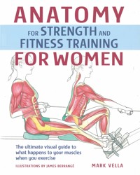
Scientific research has made it clear that a woman's body should be exercised differently from a man's, and that her training program should be adjusted to suit her body type and transformations as she moves through the various life stages.Anatomy for Strength and Fitness Training for Women is a unique guide, reference work, and graphic education tool suitable for any woman interested in understanding her own body, how it functions during exercise, and how to formulate a personal exercise program.Over 90 exercises are provided to help you sculpt your entire body with gym weights, free weights, aerobics, and stretching, including:- Swimming- Transverse activation in four-point kneeling- Barbell bench press- Cable cross-over- Freestanding barbell squats- Hip abductor machine- Seesaw with ball- Seated low cable pulley rows- Machine shoulder press- Dumbbell standing lateral raise- Rear deltoid machine- French curl- Cable tricep push-down- Supine hip flexion stretch- Plank to downward-facing dog- Child stretch- And much more!The exercises are featured in clear anatomical detail and are grouped by aerobic training, abdominals, chest, legs and hips, back and shoulders, arms, and stretches and flexibility. Each exercise incorporates a full-body illustration plus instructions on proper execution and technique. Start and finish positions are shown, and supplementary information is included by way of hints and tips.Inside this informative 144-page guide, you'll also find a method of assessing your own fitness which you can use to help you choose the correct type of program for your life stage and fitness level. Sample programs illustrate how you can choose a program based on the needs you have established in the assessment.Build a better you by understanding how your body performs when exercising, with Anatomy for Strength and Fitness Training for Women!
Sie lesen das E-Book in den Legimi-Apps auf:
Seitenzahl: 224
Das E-Book (TTS) können Sie hören im Abo „Legimi Premium” in Legimi-Apps auf:
Ähnliche


Dedication
To my late parents, I wish you could have seen this one too. This book is dedicated to you and your legacy. Time has not faded the memories, but has made them stronger.
Author’s Acknowledgments
Over the past 19 years, my journey through the health sciences has been paved with brilliant teachers and students along the way. Thank you to my colleagues Sally Lee and Tanya Wyatt, two of the best health and fitness professionals I am likely to ever know, for raising the bar. This book is a creative collaboration. To artist James Berrangé, for your enthusiasm, insight and commitment to this project – without you, this book would just be a good idea. To the team at New Holland and McGraw-Hill, thank you, thank you, thank you.
A book like this requires a lot of help. Abundant thanks are due to the following:
• For his mentorship, Professor Graham Louw, Department of Health Sciences, University of Cape Town
• For her generosity, Caroline Powrie, curator of the Anatomy Museum at Groote Schuur
• For making our lives simpler, Virgin Active Gym, Long Street, Cape Town
• Our fantastic models, Lara Turk and Terri Bruning of Cape Town City Ballet
• For her reliability, Glennis Harris of the ETA
• For their expediency and kind permission, David Brewer and Christa Dickey of the ACSM, David O’Brien and Gill Watson of Lippincott, Williams and Wilkins
Disclaimer Many of the exercises have a degree of risk of injury if done without adequate instruction and supervision. It is recommended that you get a proper fitness assessment before undertaking any of the exercises, and that you seek qualified instruction if you are a total beginner. The book does not constitute medical advice, and the author and publisher cannot be held liable for any loss, injury, or inconvenience sustained by anyone using the information in it.
CONTENTS
PART 1
ANATOMY AND EXERCISE PROGRAMMES
Building a better you
Anatomical definitions and terminology
Exercise analyses and principles
Devising a programme
Exercise programmes
PART 2
THE EXERCISES
1. Aerobic Training
Walking
Jogging and running
Aerobics
Swimming and aqua-aerobics
Aerobic machines
2. Abdominals, Stabilization and Balance
Posture basics
Seated ball balance
Transverse activation in four-point kneeling
Plank pose stabilization on ball
Shell prone ball roll-up
Abdominal stabilization programme
Two-stage crunch
Reverse incline bench sit-up
Body-weight oblique crunch – ball between legs
Combination crunch
Hip flexor apparatus
Mid-back scapular stabilization on bench
Kneeling heel touch
3. Chest
Body-weight modified push-ups
Wall push-ups on bar
Bench press machine
Incline dumb-bell bench press
Barbell bench press
Body-weight dips
Incline pec deck machine
Dumb-bell flat bench flyes
Cable cross-over
4. Legs and Hips
Squats with ball between legs
Freestanding barbell plié squats
Freestanding barbell squats
Machine incline leg press
Barbell reverse lunge
Freestanding lateral lunge
Bench step
Modified barbell bent leg deadlift
Double leg bridge with shoulder flexion
Ball bridge
Side-lying ball lift
Hip abductor machine
Hip adductor machine
Supine adductor stabilization with ball
Cable hip abductions
Prone hip extensions
Machine lying leg curl
Seesaw with ball
Yoga quad stretch with forward lean
Freestanding calf raise
Seated calf raise machine
5. Back and Shoulders
Machine cable front lateral pull-down
Chin-up assist machine
Standing cable pull-over
Standing reverse grip cable rows
Seated low cable pulley rows
Supported bent-over row machine
Dumb-bell bent-over rows
Prone back extension on ball
Back extension apparatus
Alternate arm and leg raises on ball
Machine shoulder press
Dumb-bell seated shoulder press
Dumb-bell standing lateral raise
Rear deltoid machine
Seated bent-over dumb-bell raises on ball
Rotator cuff stabilization with theraband
6. Arms
Seated overhead tricep extension on ball with theraband
Supine barbell French curl
Tricep machine
Cable tricep push-down
Tricep rope pull-down
Standing barbell curl
Seated incline dumb-bell curl with supination
Dumb-bell concentration curl
7. Stretches and Flexibility
Neck and shoulder stretch
Standing chest and anterior shoulder stretch
Ball shoulder stretch
Seated side stretch on ball
Full-body stretch
Supine hip flexion stretch
Spine roll
Side-to-side hip rolls
Supine lying gluteus stretch
Supine lying single leg hamstring stretch
Seated stride into saw stretch
Supine lying deep external rotators stretch
Standing iliopsoas stretch
Gastrocnemius stretch
Plank to downward-facing dog
Child stretch
Glossary
Index
Resources
BUILDING A BETTER YOU
Research in the last ten years has made it clear that a woman’s body should be exercised differently from a man’s. Additionally, due to the physiological differences and the distinct cycles of a woman’s life, her training programme should be adjusted to suit her body type and transformations as she moves through the various life stages. Anatomy for Strength and Fitness Training for Women combines valuable training information, exercise illustration and analysis of female-specific exercises, as well as guides on how to do each exercise properly. It is a unique guide, reference and education tool for any woman interested in understanding her own body and formulating a personal exercise programme, as well as for practitioners involved in exercise science and anatomy such as trainers, teachers and students.
How to use this book
Anatomy for Strength and Fitness Training for Women is both a visual and textual analysis of common exercises, and a guide to how to do various exercises properly.
The introductory section offers an explanation of basic anatomical movement terminology and exercise analysis, as well as a self-assessment of fitness and body types that can be used in choosing a personal exercise programme. The sample programmes that follow illustrate how to choose a routine based on the needs you have established in the assessment.
The core of the book is the exercise section. It covers aerobic training, the development of the postural stabilizers, exercises for the chest, legs and hips, back and shoulders, and arms, as well as static stretches. Each part begins with a basic introduction focused on the body part or type of training covered. Every exercise is covered independently, and defined and given some background. There is a ‘how-to’ guide to doing the exercise, as well as a visual and technical exercise analysis describing which muscles are being used as mobilizers and postural stabilizers. The start position is usually depicted in line drawings.
The adult human body has more than 600 muscles and 206 bones. In this book, emphasis is placed on the 70 or so main muscles involved in movement and stabilization. Many of the smaller muscles, as well as the deep small muscles of the spine and muscles of the hands and feet, are not given specific attention – if they were, it could take several pages to analyse just one exercise and movement.
Schematic diagram of how the exercise pages are structured
ANATOMICAL DEFINITIONS AND TERMINOLOGY
Anatomy has its own language and, although technical, it is quite logical, originating mostly from Latin and Greek root words that make it easier to learn and understand the names of muscles, bones and other anatomy parts.
The musculoskeletal system
The body comprises an integration of approximately twelve distinct systems that continuously interact to control the multitude of complex bodily functions. This book specifically illustrates and analyses the systems that control movement and posture, namely the muscular and skeletal system, which is often jointly referred to as the musculoskeletal system.
The skeletal system consists of bones, ligaments (which join bone to bone) and joints (technically known as articulations). Adults have 206 bones in a skeleton weighing about 8–9 kg (18–20 lb). The skeletal system acts as a movement framework. Muscles attach to bone and cross joints. Where they cross free-moving joints, contraction of the muscles causes joint movement.
The muscular system is made up of three types of muscle tissue, namely cardiac, smooth and skeletal.
Cardiac muscle forms the heart walls, and smooth muscle tissue is found in the walls of internal organs such as the stomach and blood vessels. Both types of muscle are activated involuntarily through the autonomic nervous system and hormonal action. Of the 700 or so muscles in a woman’s body, approximately 650 are skeletal muscles. On average, half of the body’s weight is muscle, and three-quarters of this is water.
Muscles attach to bone via tendons. These attachment points are referred to as the origin and the insertion. The origin is the point of attachment that is proximal (closest to the root of a limb), or closest to the mid-line or centre of the body. It is usually the least moveable tendon, and acts as the anchor point in muscle contraction. The insertion is the point of attachment that is distal (furthest from the root of a limb), or furthest from the mid-line or centre of the body. It is usually the most moveable part and can be drawn towards the origin.
Knowing the origin and insertion points of a muscle, which joint or joints the muscle crosses and what movement it effects at that joint or joints is a key element of exercise analysis.
Anatomical terms
The anatomical position
When learning anatomy and analysing movement we refer to the standard reference position of the human body, known as the ‘anatomical position’. All movements and the location of the anatomical structures are named as if the person was standing in this standard position.
The anatomical position is described as follows: the body is in a standing position and facing forwards, the legs and feet are together, and the arms hang loosely at the sides with the palms facing forwards.
Anatomical terms of position and direction
There are standard terms of position and direction that describe the position of body structures, or their relationship to other body parts. The human body is a complex, three-dimensional structure, and knowing the proper anatomical terms of position and direction will help you to compare one point on the body to another, and to understand where it is in relation to other anatomical structures. The terms are standard whether the person is sitting, standing or lying down.
Anatomical terms of position and direction
Position
Definition
Example of usage
Anterior
Towards the front, pertaining to the front
The abdominal muscles are found on the anterior aspect of the body
Posterior
Towards the back, pertaining to the rear
The hamstring muscles are situated on the posterior aspect of the leg
Superior
Above another structure, towards the head
The shoulder is superior to the hip
Inferior
Below another structure, towards the feet
The hip is inferior to the shoulder
Lateral
Away from the mid-line, on or towards the outside
The outside of the knee joint is its lateral aspect
Medial
Towards the mid-line of the body, pertaining to the middle or centre
The inside of the knee joint is its medial aspect
Proximal
Closest to the trunk or root of a limb. Also sometimes used to refer to the origin of a muscle.
The hip joint is proximal to the knee
Distal
Situated away from the mid-line or centre of the body, or root of a limb. Also sometimes used to refer to a point away from the origin of a muscle.
The toes are the most distal part of the leg
Superficial
Closer to the surface of the body, more towards the surface of the body than another structure
The Rectus femoris is the most superficial of the quadricep muscles
Deep
Further from the surface, relatively deeper into the body than another structure
The heart is deep to the ribs that protect it
Prone
Lying face downwards
Prone back extensions are done, as the name suggests, from a prone lying starting position
Supine
Lying on the back, face upwards
Abdominal crunches are performed from a supine lying starting position
Joint movements
Knowing and understanding the body’s movements, and at which joints they occur, is essential for analysing an exercise.
Types of joint
Some joints in the body are fixed or semi-fixed, allowing very little or no movement. For example, the bones of the skull join together in joint structures known as sutures to form fixed joints; but where the spine joins the pelvis, the sacroilliac joint is semi-fixed and allows minimal movement (‘sacro’ from sacrum, ‘iliac’ pertaining to the pelvic crest). A third category, synovial joints, comprises free-moving joints that move in different ways determined by their particular shapes, sizes and structures.
Synovial joints are the most common joints in the body. They are characterized by a joint capsule that surrounds the articulation, the inner membrane of which secretes lubricating synovial fluid, stimulated by movement. Typical synovial joints include the shoulders, knees, hips and ankles, and the joints of the hands, feet and vertebrae. Of all the joints, the knee joint is the largest, the hip joint is the strongest and the shoulder joint is potentially the most unstable.
Joint action
When performing an activity such as lifting weights or running, a combination of nerve stimulation and muscular contraction facilitates the movement that occurs at the synovial joints. When doing a bicep curl, for example, the weight rises because the angle of the elbow joint closes as the bicep muscles, which attach from the upper arm bones to the forearm (radius and ulna), shorten in contraction, thereby lifting the forearm.
Joint movement pointers
Most joint movements have common names that apply to most major joints, but there are some specific movements that only occur at one specific joint. The common joint movements occur in similar anatomical planes of motion. For example, shoulder, hip and knee flexion all occur in the same plane. This makes it more logical and easier to learn about joint movements and movement analysis. In the table on the next page, common movements are listed first, followed by specific movements that only occur at one joint.
In general, movement is paired with the joint moved, for example shoulder flexion, knee extension, spinal rotation, scapular depression and so on. Strictly speaking, it is incorrect to name the movement and a limb or body part. For example, ‘leg extension’ does not clarify where in the leg the movement occurs – at the knees, hips or ankles.
Movements generally occur in pairs. For a ‘forwards’ movement there must be a ‘backwards’ movement to return to the original starting position. Typical pairs of movements are flexion and extension, abduction and adduction, internal rotation and external rotation, and protraction and retraction. You will notice these pairs of movements when you look at the analyses in the exercise section.
All movements are named as if the person is standing in the anatomical position as described on page 9 so, for example, ‘elbow flexion’ is the same whether the person is standing, lying supine or sitting.
How a woman’s body differs from a man’s
Skeletal differences
A woman’s skeleton is usually smaller and smoother than that of a man. Overall she is 7 per cent shorter and 8 per cent smaller. However, skeletal proportions vary, so that a woman’s torso is usually longer, and proportionately longer from waist to feet. With different somatotypes (see page 14) this shifts, with ectomorphs typically having longer legs than men of the same height. This increases leverage and injury risk on the knees. A woman also has a lower centre of gravity than a man, giving her better balance.
Muscle and connective tissues
Basic measures of body strength show that females generally have 30–50 per cent less strength than males. This is mainly in the upper body, where men are about 40 per cent stronger. Women typically have less muscle mass and more body fat. Additionally, a man’s taller and wider skeletal frame provides a leverage advantage. However, ‘female muscle’ and ‘male muscle’ are exactly the same. There are no inherent gender differences in muscle quality or capacity, and women can generally generate the same force per unit of muscle as men. Furthermore, with training they make the same relative strength improvements.
In certain sports, such as climbing, dance and aspects of gymnastics, the lower centre of gravity, flexibility, strength-to-weight ratio, and shorter levers give a woman a better relative strength ratio.
Fat tissue
Two types of body fat make up total body fat, namely fat stored mainly within the organs and muscles essential for the various body processes, and adipose tissue stored more underneath the skin of the body. Excess fat is stored in adipose tissue.
High testosterone and growth hormone in men creates greater muscle mass with higher basal metabolism-consuming energy. Men can therefore eat relatively more and expend more energy than women. Oestrogen in women, on the other hand, increases fat storage in a woman’s body.
Women have more essential body fat than men (3 per cent as opposed to 12 per cent), as well as greater body fat percentages. For a woman in her twenties a healthy body fat percentage would be 23–27 per cent. The equivalent in a man would be 16 per cent. After the age of 45, a healthy woman would have 32 per cent body fat compared with 25 per cent in a man.
In men, adipose fat deposition occurs mainly around the stomach. In women it is distributed between the hips and buttocks, in the inner thighs and in small sites at the backs of the upper arms and around the navel and medial knee. The breasts are also filled with fat, which encloses the mammary glands.
Women are more likely to experience weight shifts than men, particularly if they experience early menopause, or are sedentary or overweight to begin with.
Elevated fat levels are therefore a normal part of a woman’s physiology. Exercise consultation should define excess versus healthy fat levels, and programming should set realistic goals and expectations of what is possible and healthy to strive for.
Major joint movements
GENERAL MOVEMENTS
Plane
Description
Example
Abduction
Coronal
Movement away from the mid-line
Hip abduction
Adduction
Coronal
Movement towards the mid-line
Hip adduction
Flexion
Sagittal
Decreasing the angle between two structures
Moving the forearm towards the upper arm. Standing Barbell Curl
Extension
Sagittal
Increasing the angle between two structures
Moving the forearm away from the upper arm. As above, downward phase.
Medial rotation (internal rotation)
Transverse
Turning around the vertical axis of a bone towards the mid-line
Cable Cross-over
Lateral rotation (external/outward rotation)
Transverse
Turning around the vertical axis of a bone away from the mid-line
Turning at the waist
Circumduction
All planes
Complete circular movement at shoulder or hip joints
Swinging the arms in circles
SPECIFIC MOVEMENTS
1. Ankle movements
Plantarflexion
Sagittal
Moving the foot downwards
Freestanding Calf Raise (upward phase)
Dorsiflexion (Dorsal flexion)
Sagittal
Moving the foot towards the shin
Freestanding Standing Calf Raise (downward phase)
2. Forearm movements (the radioulnar joint)
Pronation
Transverse
Rotating the wrist and hand medially from the elbow
Standing Dumb-bell Curl
Supination
Transverse
Rotating the wrist and hand laterally from the elbow
Making circular movements with your hand
3. Scapula movements
Depression
Coronal
Movement of the scapulae inferiorly, e.g. squeezing scapulae downwards
To stabilize the shoulder girdle, e.g. Hip Flexor Apparatus
Elevation
Coronal
Movement of the scapulae superiorly, e.g. hunching the shoulders
Dumb-bell Seated Shoulder Press (up)
Abduction (protraction)
Transverse
Movement of the scapulae away from the spine
Seated Low Cable Pulley Rows
Adduction (retraction)
Transverse
Movement of the scapulae towards the spine
As above
Downward rotation (medial rotation)
Coronal
Scapulae rotate downwards, in the return from upward rotation
Machine Cable Front Lateral Pull-down
Upward rotation (lateral rotation)
Coronal
Scapulae rotate upwards. The inferior borders of the scapulae move laterally and upwards.
As above
4. Shoulder movements
Horizontal abduction/extension (transverse abduction)
Transverse
Movement of the humerus across the body away from the mid-line
Dumb-bell Flat Bench Flyes
Horizontal adduction/flexion (transverse adduction)
Transverse
Movement of the humerus across the body towards the mid-line
As above
5. Spine/trunk movements
Lateral flexion
Coronal
Movement of the trunk away from the mid-line in the coronal plane
Seated Side Stretch on Ball
EXERCISE ANALYSES AND PRINCIPLES
Purpose of exercise analysis
Analysing an exercise enables you to understand what joints and muscles are being used in a certain movement and how they are moving. Changing the movement, or doing the movement incorrectly, affects both the muscles used to perform the exercise and how they are used. Exercise analysis can also help you to determine if the muscles you intend to train are in fact being utilized in the specific exercise being done, as well as whether you are doing the exercise correctly.
Somatotyping
The word ‘somatotyping’ literally means ‘body typing’. The term is used in a system of classification of human physical types, according to which human beings can be grouped in terms of three extreme body types. Endomorphs tend to relative fatness, mesomorphs to relative musculoskeletal robustness or muscularity, and ectomorphs to relative linearity or slenderness. Assessing your somatotype will indicate what your particular mix of these three types is. In turn, the somatotype will suggest certain traits – for example, ectomorphs often show better aerobic ability than endomorphs, who show better power and strength capacity.
Endomorphs
Female endomorphs tend to be pear shaped (which means that they carry weight mainly around the hips), and convert to more of an apple shape after the menopause. This is due to increased intra-abdominal fat being laid down in the abdomen at this time. Male endomorphs tend to be apple shaped, that is abdominally obese. Overall, endomorphs struggle to lose weight and tend to gain it more easily than the other somatotypes. Women are more likely to be endomorphic than men.
Mesomorphs
Female mesomorphs tend to be hourglass shaped, with smaller waists compared with the bust and hips, which are the same size. This shape, although popular, is less common in women than in men.
Ectomorphs
Ectomorphic women struggle to gain weight and have less curvature and shape than the other somatypes. While they recover relatively quickly from exercise, their longer levers and reduced muscle mass increase their need for postural and strength conditioning.
Starting out
As you kindle the exercise habit, expect to go through three distinct phases. Knowing these will help put your feelings in each phase into perspective. It is quite normal for progress to seldom be smooth from one phase to another. It is common to have lapses and relapses, but with consistent perseverance you will move through them. If you get bored easily, introduce some variation to your programme. It may also be an idea to initially train with an exercise partner.
The three phases of motivation
Resistance phase
In this initial phase, which lasts 6–12 weeks, expect your motivation to be relatively low and your resistance to exercise to be high. The key is to be consistent. Keep your workout appointments. The programme should be fairly uncomplicated and err on the conservative side, but challenging enough to feel worthwhile. This phase often reveals what the assessment did not, and may lead to you reconsidering your goals or choice of programme. While you are probably not seeing much physical change, you should be feeling improvements by the third week. Most people report better sleep as the first change.
Transition phase
In the second phase, which lasts some 3–6 months, physical results should have started to show. You may start to over-train because you are tempted to try for more changes. Instead, keep trying to get maximum results from minimum effort. Use your three-month fitness reassessment to make necessary adjustments in your programme based on how your test scores have changed. You should also begin to become more aware of the subtler aspects of your fitness and health. Training frequency usually reaches its peak by this stage.
Intrinsic phase
This point is reached when your motivation to exercise comes from the inside and is an established lifestyle habit. You may by now have homed in on some aspect of training you enjoy, such as aerobics or swimming, and be including exercise as an established part of your diary. To offset a training plateau and getting into a complete rut, try to keep varying the programme every three months or so. This may also be a good time to consult a specialist trainer to further individuate your training or acquire more technical information and skills.
Principles you should know
Functional training involves using your body in a way that is similar to day-today movement. Most functional training employs compound exercises, namely those that use more than one muscle group and joint at a time, and that are generally close chain and weight bearing. Functional training is a part of health-related fitness, which is a part of everyday health, ensuring optimum quality of life for everyday demands. It is usually defined by the variables of cardiovascular fitness, flexibility, strength, strength endurance and flexibility, with cardiovascular fitness being regarded as the most important because it has the greatest influence on our health. The definition has begun to also consider and include aspects such as postural strength.
The principles of exercise are based on the laws of nature. Understanding them will help you exercise in a safe and effective manner, and ensure the best results.
SAID principle
According to the principle of Specific Adaptation to Imposed Demands (SAID), your body will predictably change, through an adaptive process, in response to the exercise demands that are placed on it. This means that if you stretch regularly (stimulus) it can improve your flexibility (response) as your body becomes more supple (adaptation) to handle the increased range of movement demands placed upon it. Each component of physical fitness can be developed and trained. The adaptation is also specific to the response. If you run regularly, you will become a better runner. If you cycle regularly, you will become a better cyclist. Your training programme should thus be specific to the desired response.
This principle also illustrates why it is important to train in postures and functional positions, and in a manner that is closest to the demands of your life. For example, exercising in standing positions contributes to strengthening the spine and trunk and leg stabilizers, contributing to their fitness for everyday activities. How fast adaptation will occur varies in individuals, but those just beginning to exercise or in the lowest ranges of functional capacity are likely to improve the quickest.
Overload principle
In order to gain improvement in fitness, the body must be stimulated beyond its current capacity. Therefore if you wish to gain strength, you must train with a weight stimulus that is higher than your current capacity. This amount of stimulus is called the overload. Too great an overload can, however, cause injury.
FITT principle
There are four major variables of overload to any training plan or exercise prescription. These are frequency, intensity, type and time or duration. Your training programme will be incomplete if these are not incorporated into your plan. To maximize overload you can increase training in any of the four major variables.
Progression principle
Over time, adaptation to the initial training stimulus occurs. To overcome this plateau, the overload must further increase.
Reversibility principle
It is often said, ‘Use it or lose it.’ This is a simple illustration of the reversibility principle. Adaptation made through training stimulus may be lost if the stimulus is discontinued.
Rest/effort balance principle
Rest and recovery periods are as much a part of training as is training itself, and must be balanced with training. Resting too much results in regression, whereas training too much results in overtraining.


