151,99 €
Mehr erfahren.
- Herausgeber: John Wiley & Sons
- Kategorie: Fachliteratur
- Sprache: Englisch
The current state of knowledge on this increasingly important subject is beautifully described in this, the first truly comprehensive text of allergic diseases affecting the major veterinary species. It will be an invaluable guide to students, clinicians and researchers alike. From the Foreword by Professor Richard Halliwell, MS, PhD, VetMB, MRCVS, Dip ACVD
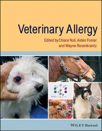
Veterinary Allergy is the first comprehensive, high quality reference dealing with all aspects of veterinary allergy in all species and all body systems involved with allergy. Providing solid breadth and excellent depth of coverage, it deals with the immunopathology of the various allergic conditions as well as with clinical presentation, diagnosis and treatment of veterinary allergic diseases.
Key features:
- Broad species coverage, organised by sections on dogs, cats, horses, and other domestic species including large animals, birds and small mammals
- Provides details on diseases affecting the skin, respiratory tract and gut
- Contains practical information for clinicians on management of allergic diseases
- Includes fully-referenced high level detail suitable for specialists and researchers
- Focused on evidence-based medicine and science
- The editors have extensive experience and are respected as world-wide experts in the field
- Every chapter is written by an expert in that particular topic
Anyone seeking information and references on any aspect of allergic conditions in any species should find this book helpful. The book will be of interest to clinicians in first opinion practice, specialists in veterinary dermatology, gastroenterology, internal medicine, and immunology as well as specialists-in-training in those fields and veterinary students.
The focus on underlying principles and practical clinical aspects is admirable. Immediately useful for practical utilization by practicing clinicians. Dr. Andrew Mackin BSc BVMS MVS DVSc FACVSc DSAM, Diplomate, American College of Veterinary Internal Medicine, Professor and Service Chief, Small Animal Internal Medicine, Dr. Hugh G. Ward Endowed Chair of Small Animal Veterinary Medicine, College of Veterinary Medicine, Mississippi State University, USA
Sie lesen das E-Book in den Legimi-Apps auf:
Seitenzahl: 1326
Veröffentlichungsjahr: 2013
Ähnliche


Table of Contents
Dedication
Title page
Copyright page
Acknowledgments
Contributors
Foreword
Introduction: the immunological basis of allergic diseases
Introduction
The multifactorial nature of allergy
Future progress
Part 1: Canine Allergy
Section 1: Canine Atopic Dermatitis
1: Introduction: canine atopic dermatitis as an evolving, multifactorial disease
2: Canine immunoglobulin E
Introduction
The role of immunoglobulin E in atopic disease
Immunoglobulin E interaction with immune and inflammatory cells
The immunoglobulin E network of soluble receptors and autoantibody complexes
Therapeutic anti-immunoglobulin E
Conclusion
3: The aberrant immune system in atopic dermatitis
Atopic dermatitis as a type I hypersensitivity: the role of mast cells, histamine, IgE, and leukotrienes
The role of dendritic cells
T cells: different populations and cytokine response
Summary of proposed pathogenesis
4: Allergens and environmental influence
Introduction
Sources of allergens
Environment and canine atopic dermatitis
Allergen specificities
Conclusion
5: The genetics of canine atopic dermatitis
Introduction
Investigating atopic dermatitis-associated genotypes
Genomic studies in canine atopic dermatitis
Conclusion
Glossary
6: Skin barrier and its role in the pathophysiology of atopic dermatitis
Introduction
Methods to determine impairment of barrier function of the canine skin
Extracellular lipids in the stratum corneum and atopic dermatitis
Current evidence for aberrant filaggrin expression in dogs with atopic dermatitis
Other barrier abnormalities in human atopic dermatitis
Future perspectives for the investigation of cutaneous barrier function in canine atopic dermatitis
7: The role of bacterial agents in the pathogenesis of canine atopic dermatitis
Introduction
Environmental exposure
The defective skin barrier
Bacterial virulence and the pathogenesis of canine pyoderma
Conclusion
8: The role of fungal agents in atopic dermatitis
Introduction
Overview of fungi in human and canine atopic dermatitis
Environmental fungal allergens in human atopic dermatitis
Environmental fungal allergens in canine atopic dermatitis
Dermatophytosis in human and canine atopic dermatitis
Malassezia yeasts in human atopic dermatitis
Malassezia yeasts in canine atopic dermatitis
Conclusion
9: Clinical signs of canine atopic dermatitis
Signalment of CAD dogs
History of canine atopic dermatitis dogs
Clinical signs of canine atopic dermatitis
Breed-associated phenotypes
10: Diagnosis of canine atopic dermatitis
The approach to the chronic pruritic dog
Establishing the diagnosis of CAD
Determining environmental allergens for CAD
Conclusion
11: Allergen avoidance
The role of indoor allergens
The role of outdoor allergens
Conclusion
12: Allergen-specific immunotherapy
Introduction
Mechanism of action
Selection of allergens
Conventional allergen-specific immunotherapy
Rush immunotherapy
Intralymphatic immunotherapy
Sublingual immunotherapy
Allergen-specific immunotherapy and concurrent drug therapy
13: Guidelines for symptomatic medical treatment of canine atopic dermatitis
An integrated approach to treatment
Managing acute flares of canine atopic dermatitis
Long-term management of chronic atopic dermatitis
14: Non-conventional treatments
Introduction
Pentoxifylline
Tacrolimus
Misoprostol
Chinese herbal medicine
Capsaicin
Leukotriene inhibitors
Serotonin uptake inhibitors and tricyclic antidepressants
Probiotics
Therapy with helminths
Interferon-γ
Hardy kiwi preparation
Heat-killed Mycobacterium vaccae
Conclusion
Section 2: Food Hypersensitivity
15: The pathogenesis of food allergy
Introduction: lessons from human food allergy
What do we know about canine food allergy?
Conclusion
16: Cutaneous manifestations of food hypersensitivity
Historical perspective
Epidemiology
Sex, age (at onset), and breed predilection
Pruritus
Lesions and pattern of distribution
Otitis externa
Secondary skin diseases
Particular and rare dermatological signs
Gastrointestinal signs in dogs with skin disease
Other non-dermatological signs associated with skin disease
17: Adverse reactions to food: a gastroenterologist's perspective
Adverse food reactions
Epidemiology of adverse food reactions
Clinical presentation
Differential diagnoses
Diagnosis
18: Diagnostic workup of food hypersensitivity
Hypoallergenic diets
Invivo tests: intradermal testing and gastroscopy
Invitro tests
19: Long-term management of food hypersensitivity in the dog
Abbreviations
Introduction
Pragmatic considerations about diets for food hypersensitivity
Non-allergen aspects of the diet associated with clinical improvement
Alternative non-dietary treatments of food hypersensitivity
Sensitization to novel proteins
Eventual restoration of tolerance to allergens
Conclusions and recommendations
Section 3: Flea Bite Allergy
20: Flea biology and ecology
Introduction
Morphology
Distribution
Life cycle
Veterinary importance
Conclusion
21: The pathogenesis of flea bite allergy in dogs
Introduction
The flea allergen
The immunopathogenesis of FAD
Conclusion
22: Clinical signs of flea allergy dermatitis in dogs
Introduction
Is flea infestation easy to diagnose?
Is there a season for flea allergy dermatitis?
No epidemiological clues!
‘Classical lesions’ of flea allergy dermatitis
Pyotraumatic dermatitis
Urticaria
Atopic dermatitis flares
Fibropruritic nodules
Microbial infections
Differential diagnoses
23: Diagnostic investigation of canine flea bite allergy
Live flea challenge test
Intradermal testing
Serological testing
Histopathology
Conclusion
24: Implementing an effective flea control programme
Introduction
General control strategies and integrated flea control
Perceptions of reduced efficacy
Conclusion
25: Symptomatic relief for canine flea bite hypersensitivity
Antimicrobial agents
Glucocorticoids
Other systemic antipruritic drugs
Topical therapy
Immunotherapy
Conclusion
Section 4: Complicating Infections in Allergic Dogs
26: Complicating microbial skin infections in allergic dogs
Importance and prevalence
Pathogens
Clinical manifestations
Diagnostic approach
Therapy
Prevention
27: Otitis in the allergic dog
Introduction
The relationship of otitis to allergy
Clinical features of otitis in allergy
Management of otitis in allergic patients
Conclusion
Section 5: Other Allergic Diseases in Dogs
28: Contact allergy
Is it a rare disease?
Causes of contact allergy
Pathogenesis
Clinical presentation
Diagnosis
Management
29: Venomous insect hypersensitivity
Introduction
Pathogenesis
Treatment
30: Canine urticaria and angioedema
Introduction
Aetiology
Pathogenesis
History, clinical signs, and differential diagnosis
Diagnostic evaluation
Treatment
Part 2: Feline Allergy
Section 1: Cutaneous Allergy in Cats
31: Pathogenesis—immunopathogenesis
Genetic background
Feline skin barrier and exposure to allergens
Antigen-specific immunoglobulin E
Dendritic cells
T cells
Eosinophils
Mast cells
Role of microbial factors
32: Clinical presentations and specificity of feline manifestations of cutaneous allergies
Classical presentations of feline atopic-like disease and cutaneous adverse reactions to food: eosinophilic dermatitis, miliary dermatitis, self-induced alopecia, and head and neck pruritus
Signalment and history of cats with atopic-like disease and/or cutaneous adverse reactions to food
Classical presentations of atopic-like disease and cutaneous adverse reactions to food
Distribution patterns
Other signs
Specificity of reaction patterns
The specificity of feline manifestations of cutaneous allergy
33: Complications of cutaneous skin allergies (skin infections)
Introduction
Superficial bacterial infections
Superficial fungal infections
Viral infections
34: Diagnostic investigation of the allergic feline
Introduction
Key aspects of the history
Environmental allergy
35: Symptomatic treatments
Managing secondary pruritic conditions
Allergen avoidance
Medical management of cutaneous allergy
36: Allergen-specific immunotherapy
Principles and considerations for allergen-specific immunotherapy
Options for allergen-specific immunotherapy
Section 2: Feline Asthma
37: Feline asthma
Introduction
Immunopathology
Clinical presentation
Diagnosis
Treatment
Section 3: Flea Bite Allergy
38: Pathogenesis
Flea saliva
Allergenicity of flea saliva
Immune responses to flea saliva in cats and other species
39: Clinical presentations
Miliary dermatitis
Symmetrical alopecia
Head and neck excoriations
Eosinophilic granuloma complex
Other signs
40: Diagnostic workup
Live flea challenge test
Intradermal testing
Serological testing
Functional in vitro test
Histopathology
41: Therapy
Introduction
Main difficulties and pitfalls
Active ingredients and formulations
Recommendations
Section 4: Mosquito Bite Allergy
42: Mosquito bite
Pathogenesis
Clinical features
Diagnostic tests
Treatment
Part 3: Equine Allergy
Section 1: Culicoides Hypersensitivity and Other Insect Allergies
43: Pathogenesis and epidemiology of Culicoides hypersensitivity
Introduction
Epidemiology
Culicoides characteristics, life cycle, and breeding sites
Pathogenesis
Culicoides allergens
44: Equine immunoglobulin E
Structure and function of IgE
Antibodies for detection of equine IgE
IgE in Culicoides hypersensitivity
Conclusion
45: Clinical manifestations of Culicoides hypersensitivity
46: Culicoides hypersensitivity: diagnosis
Introduction
Allergy testing
Serum testing
Intradermal testing
Allergens sources
47: Culicoides hypersensitivity: therapy
Prevention of Culicoides hypersensitivity
Vaccinations
Environmental control
Insect control
Kairomones
Topical non-insecticide therapy
Systemic therapy
Conclusion
48: Other biting insect allergies
Introduction
Allergen material sources and characterization
Pathogenesis of biting insect hypersensitivity
Clinical signs
Differential diagnosis
Diagnosis
Diagnostic confirmation
Treatment
Selected biting Diptera
Section 2: Atopic Disease in Horses—Atopic Dermatitis and Food Hypersensitivity
49: Equine atopic dermatitis: pathogenesis
Defining atopic dermatitis
The evidence for atopic dermatitis in horses
Food allergy in horses as part of atopic diathesis
50: Clinical aspects of equine atopic disease
Introduction
History and signalment
Clinical lesions
Concurrent diseases
51: Equine urticaria
Overview
Mast cell degranulation
Acute versus chronic urticaria
Potential causes for and recommended approach to chronic or recurrent urticaria
Treatment of urticaria
52: Equine headshaking syndrome
Clinical syndrome
Causes of headshaking syndrome
53: Diagnostic workup of equine atopic disease
Introduction
Dermatopathology
IgE testing
Serum invitro testing
Intradermal testing
Adverse food reaction testing
54: Equine atopic disease symptomatic therapy and allergen-specific immunotherapy
Introduction
Avoidance
Elimination diet trials
Topical therapy
Systemic therapy
Section 3: Recurrent Airway Obstruction and Inflammatory Airway Disease
55: Recurrent airway obstruction and inflammatory airway disease
Pathogenesis
Clinical aspects and diagnosis
Further diagnostic investigation
Therapy
Section 4: Contact and Other Allergic Diseases
56: Equine allergic contact dermatitis
Pathogenesis
Clinical features
Diagnosis
Photohypersensitivity
Part 4: Allergy in Other Domestic Species
57: Immunopathogenesis of allergic skin disease in livestock
Introduction
The innate response
The adaptive response
58: Psoroptes ovis
Incidence and importance
Clinical signs
Pathology of primary infestations
Challenge infestations
Overview, including molecular aspects, of the immunopathogenesis of sheep scab
Diagnosis
Treatment and prevention
59: Allergic diseases of livestock species
Introduction
Camelids
Cattle
Goats
Pigs
Sheep
Flea bite hypersensitivity in livestock species
60: Allergies in birds
Feather-picking
Allergen testing in avian species
Treatment options
61: Allergic diseases in other pets (rodents, rabbits, and ferrets)
Respiratory disease
Gastrointestinal disease
Dermatological disease
Index
In memory of my dear colleague and friend Stefano Toma, who is greatly missed. (CN)
I dedicate this book to Susan Shaw in recognition of her great enthusiasm and ability to share with me, as for many vets, her considerable knowledge and understanding of veterinary medicine and dermatology. (AF)
To my cherished friend and mentor, Peter Ihrke, who first introduced me to this wonderful specialty and continues to be an inspiration to all. (WR)
This edition first published 2014
© 2014 by John Wiley & Sons, Ltd
Registered office: John Wiley & Sons, Ltd, The Atrium, Southern Gate, Chichester, West Sussex, PO19 8SQ, UK
Editorial offices: 9600 Garsington Road, Oxford, OX4 2DQ, UK
The Atrium, Southern Gate, Chichester, West Sussex, PO19 8SQ, UK
111 River Street, Hoboken, NJ 07030-5774, USA
For details of our global editorial offices, for customer services and for information about how to apply for permission to reuse the copyright material in this book please see our website at www.wiley.com/wiley-blackwell
The right of the author to be identified as the author of this work has been asserted in accordance with the UK Copyright, Designs and Patents Act 1988.
All rights reserved. No part of this publication may be reproduced, stored in a retrieval system, or transmitted, in any form or by any means, electronic, mechanical, photocopying, recording or otherwise, except as permitted by the UK Copyright, Designs and Patents Act 1988, without the prior permission of the publisher.
Designations used by companies to distinguish their products are often claimed as trademarks. All brand names and product names used in this book are trade names, service marks, trademarks or registered trademarks of their respective owners. The publisher is not associated with any product or vendor mentioned in this book. It is sold on the understanding that the publisher is not engaged in rendering professional services. If professional advice or other expert assistance is required, the services of a competent professional should be sought.
The contents of this work are intended to further general scientific research, understanding, and discussion only and are not intended and should not be relied upon as recommending or promoting a specific method, diagnosis, or treatment by health science practitioners for any particular patient. The publisher and the author make no representations or warranties with respect to the accuracy or completeness of the contents of this work and specifically disclaim all warranties, including without limitation any implied warranties of fitness for a particular purpose. In view of ongoing research, equipment modifications, changes in governmental regulations, and the constant flow of information relating to the use of medicines, equipment, and devices, the reader is urged to review and evaluate the information provided in the package insert or instructions for each medicine, equipment, or device for, among other things, any changes in the instructions or indication of usage and for added warnings and precautions. Readers should consult with a specialist where appropriate. The fact that an organization or Website is referred to in this work as a citation and/or a potential source of further information does not mean that the author or the publisher endorses the information the organization or Website may provide or recommendations it may make. Further, readers should be aware that Internet Websites listed in this work may have changed or disappeared between when this work was written and when it is read. No warranty may be created or extended by any promotional statements for this work. Neither the publisher nor the author shall be liable for any damages arising herefrom.
Library of Congress Cataloging-in-Publication Data
Veterinary allergy / edited by Chiara Noli, Aiden Foster, Wayne Rosenkrantz.
pages cm.
Includes bibliographical references and index.
ISBN 978-0-470-67241-9 (pbk. : alk. paper) – ISBN 978-1-118-73881-8 – ISBN 978-1-118-73889-4 (epub) – ISBN 978-1-118-73890-0 (emobi) – ISBN 978-1-118-73891-7 (epdf) 1. Allergy in dogs. 2. Allergy in cats. 3. Allergy in animals. I. Noli, Chiara, editor of compilation. II. Foster, Aiden P., editor of compilation. III. Rosenkrantz, Wayne, editor of compilation.
[DNLM: 1. Hypersensitivity–veterinary. 2. Allergens–immunology. 3. Dermatitis–veterinary. 4. Immune System Diseases–veterinary. 5. Veterinary Medicine–methods. SF 757.2]
SF992.A44V48 2014
636.089'6975–dc23
2013026498
A catalogue record for this book is available from the British Library.
Wiley also publishes its books in a variety of electronic formats. Some content that appears in print may not be available in electronic books.
Cover images: dog and horse – courtesy of Wayne Rosenkrantz; alpaca – courtesy of Natalie Jewell and the AHVLA library, Crown Copyright 2013; cat – courtesy of Karen Clifford; centre image – courtesy of Valerie A. Fadok
Cover design by Steve Thompson
Acknowledgments
We are grateful to Wiley for taking on this publishing project and for bringing it to fruition so quickly and efficiently. We are particularly grateful to Rupert Cousens, Jessica Evans and Nancy Turner for setting up and running the project; to Elizabeth Paul and Ruth Swan for sorting out the production issues including copyediting and typesetting.
A book project of this size is time consuming for all concerned and as editors we have been delighted at the speed and enthusiasm of authors to provide their chapters and agree to revisions and changes. Inevitably the editing has taken us away from our families and everyday work, and we are grateful to our spouses for their support and understanding and to our colleagues for their patience and advice.
It has taken three years to bring the original concept to print, and we hope that readers will find this book useful; as editors it has been an enjoyable learning experience, working together and with the authors.
Contributors
Emmanuel Bensignor
Dermatology Referral Practice
Rennes-Cesson
France
Kerstin Bergvall
Swedish University of Agricultural Sciences
Department of Clinical Sciences
Uppsala
Sweden
Ross Bond
Department of Clinical Sciences and Services
Royal Veterinary College
Hatfield
UK
Mona J. Boord
Animal Dermatology Clinic
San Diego, CA
USA
Stewart T.G. Burgess
Moredun Research Institute
Pentlands Science Park
Edinburgh, Midlothian
UK
Marie-Christine Cadiergues
Toulouse Veterinary School
Toulouse
France
Didier N. Carlotti
Service de Dermatologie
Aquivet Clinique Vétérinaire
Eysines, Bordeaux
France
Nick J. Cave
Institute of Veterinary, Animal and Biomedical Sciences
Massey University
Palmerston North
New Zealand
Michael J. Day
School of Veterinary Sciences
University of Bristol
Langford, North Somerset
UK
Douglas J. DeBoer
Department of Medical Sciences
School of Veterinary Medicine
University of Wisconsin–Madison
Madison, WI
USA
Alison B. Diesel
Department of Small Animal Clinical Sciences
College of Veterinary Medicine and Biomedical Sciences
Texas A&M University
College Station, TX
USA
Michael W. Dryden
Department of Diagnostic Medicine and Pathobiology
Kansas State University
Manhattan, KS
USA
Valerie A. Fadok
North Houston Veterinary Specialists
Spring, TX
USA
Claude Favrot
Vetsuisse Faculty
University of Zurich
Zurich
Switzerland
Aiden P. Foster
Animal Health and Veterinary Laboratories Agency
Shrewsbury
Shropshire
UK
Vinzenz Gerber
Swiss Institute of Equine Medicine (ISME)
Vetsuisse Faculty
University of Bern
and ALP-Haras Avenches
Switzerland
Paola Gianella
Dipartimento di Scienze Veterinarie
Università degli Studi di Torino
Grugliasco
Italy
Craig E. Griffin
Animal Dermatology Clinic
San Diego, CA
USA
Joya Griffin
Animal Dermatology Clinic
Louisville, KY
USA
Richard E.W. Halliwell
Royal (Dick) School of Veterinary Studies
University of Edinburgh
Easter Bush Veterinary Centre
Roslin, Midlothian
UK
Bruce Hammerberg
Center for Comparative Medicine and Translational Research
College of Veterinary Medicine
North Carolina State University
Raleigh, NC
USA
Eman Hamza
Clinical Immunology Group
Department of Clinical Research and Veterinary Public Health
Vetsuisse Faculty
University of Bern
Bern
Switzerland
Peter Hill
Companion Animal Health Centre
School of Animal and Veterinary Sciences
University of Adelaide
Roseworthy, SA
Australia
Hilary A. Jackson
Dermatology Referral Service Ltd
Glasgow
UK
Janet D. Littlewood
Veterinary Dermatology Referrals
Landbeach, Cambridge
UK
David H. Lloyd
Department of Clinical Sciences and Services
Royal Veterinary College
Hatfield
UK
Anette Loeffler
Royal Veterinary College
University of London
London
UK
Dawn Logas
Veterinary Dermatology Center
Maitland, FL
USA
Gwendolen Lorch
The Ohio State University
College of Veterinary Medicine
Columbus, OH
USA
Rosanna Marsella
Department of Small Animal Clinical Sciences
College of Veterinary Medicine
University of Florida
Gainesville, FL
USA
Eliane Marti
Clinical Immunology Group
Department of Clinical Research and Veterinary Public Health
Vetsuisse Faculty
University of Bern
Bern
Switzerland
Karen A. Moriello
Department of Medical Sciences
School of Veterinary Medicine
University of Wisconsin–Madison
Madison, WI
USA
Daniel O. Morris
School of Veterinary Medicine
University of Pennsylvania
Philadelphia, PA
USA
Ralf S. Mueller
Centre for Clinical Veterinary Medicine
Ludwig Maximilian University
Munich
Germany
Masahiko Nagata
ASC Dermatology Service
Tokyo;
Synergy Animal General Hospital Dermatology Service
Saitama
Japan
Claudia S. Nett-Mettler
Vetderm.ch-Dermatologie und Allergologie fuer Tiere
c/o Ennetsee Klinik für Kleintiere AG
Huenenberg
Switzerland
Koji Nishifuji
Department of Veterinary Medicine
Tokyo University of Agriculture and Technology
Fuchu, Tokyo
Japan
Chiara Noli
Servizi Dermatologici Veterinari
Peveragno
Italy
James O. Noxon
Hixson-Lied Small Animal Hospital
Department of Veterinary Clinical Sciences
College of Veterinary Medicine
Iowa State University
Ames, IA
USA
Tim Nuttall
Royal (Dick) School of Veterinary Studies
University of Edinburgh
Easter Bush Veterinary Centre
Roslin, Midlothian
UK
William E. Oldenhoff
Department of Medical Sciences
School of Veterinary Medicine
University of Wisconsin–Madison
Madison, WI
USA
Laura Ordeix
Department of Animal Medicine and Surgery
Veterinary School
Autonomous University of Barcelona;
Dermatology Service
Hospital Ars Veterinaria
Barcelona
Spain
Annette D. Petersen
Department of Small Animal Clinical Sciences
College of Veterinary Medicine
Michigan State University
East Lansing, MI
USA
Pascal Prélaud
Clinique Advetia
Paris
France
Chris Reeder
Animal Dermatology Clinic
Louisville, KY
USA
Carol R. Reinero
Department of Veterinary Medicine and Surgery
University of Missouri
Columbia, MO
USA
Petra J. Roosje
Division of Clinical Dermatology
Department of Clinical Veterinary Medicine
Vetsuisse Faculty
University of Bern
Bern
Switzerland
Wayne Rosenkrantz
Animal Dermatology Clinic
Tustin, CA
USA
Edmund J. Rosser Jr
Department of Small Animal Clinical Sciences
College of Veterinary Medicine
Michigan State University
East Lansing, MI
USA
Ian Sayers
South Devon Exotics
Silverton Veterinary Practice Ltd
Paignton, Devon
UK
Harold C. Schott II
Department of Large Animal Clinical Sciences
College of Veterinary Medicine
Michigan State University
East Lansing, MI
USA
Adri van den Broek
Royal (Dick) School of Veterinary Studies
University of Edinburgh
Easter Bush Veterinary Centre
Roslin, Midlothian
UK
Bettina Wagner
College of Veterinary Medicine
Cornell University
Ithaca, NY
USA
Stephen White
School of Veterinary Medicine
University of California
Davis, CA
USA
Anthony A. Yu
Yu of Guelph Veterinary Dermatology
Guelph Veterinary Specialty Hospital
Guelph, ON
Canada
Foreword
The term ‘allergy’ was introduced by the Austrian physician Clemens von Pirquet in 1906 [1], however with a somewhat different meaning to that of today. He was studying the immune response to tuberculosis and diphtheria, and was thus working at the interface between immunity and hypersensitivity. He proposed the use of the term ‘allergy’ to imply ‘altered reactivity’ in the host. Thus allergy was not a disease per se, but rather a state that would result in a hypersensitivity reaction if appropriately challenged. This concept was gradually discarded despite some attempts to keep it alive. Tremendous advances in the understanding of the science behind allergy in man—now used synonymously with hypersensitivity, were made between 1920 and 1940. Notable were the studies by Prausnitz and Küstner [2] who described the skin-sensitizing antibody that was responsible for many allergic reactions. Then in the 1930s Coca [3] introduced the term ‘atopy’ which was derived from the Greek and translated literally as ‘strange disease’, to encompass the triad of the familial diseases of allergic asthma, allergic rhinitis, and atopic dermatitis. They also applied the term ‘reagin’ to the skin-sensitizing antibody of Prausnitz and Küstner. Anyone who reads these early publications cannot but marvel at the painstaking and insightful work, undertaken without the aid of modern-day techniques and at the generally sound conclusions that were reached. The next major step forward was the demonstration by Ishizaka and colleagues that the reagin belonged to a hitherto undescribed antibody class that they termed ‘IgE’ [4].
Over the years, a wide range of diseases of man mediated by diverse immunological mechanisms were described that could be ascribed to hypersensitivity reactions; Gell and Coombs believed that it was necessary to introduce a system of classification [5]. They proposed four categories, namely Type 1 hypersensitivity (IgE mediated), Type 2 (cytotoxic), Type 3 (immune complex), and Type 4 (cell-mediated). Robin Coombs was in fact a veterinarian, and although he never practised, he was responsible for training a number of veterinary immunologists who passed through his laboratory in Cambridge. However useful this classification undoubtedly was, it has become clear that few allergic diseases are caused exclusively by one type of hypersensitivity and most result from a combination. In the last three decades, and aided by the advent of molecular biological techniques, the science of allergy has advanced exponentially to become a highly sophisticated science and a major branch of human medicine.
In contrast, veterinary allergy (now defined as ‘a hypersensitivity reaction initiated by a specific immunological response to an allergen and mediated by antibodies or cells’ [6]) was slower to emerge as a recognized discipline. In large measure this can be ascribed to fewer resources for research, but also to the fact that we are concerned with multiple species, each one of which requires the development of species-specific reagents. And, of course, no formal specialist status exists for the discipline in any country. Nonetheless, its importance in everyday veterinary practice is unquestioned—indeed it is unlikely that a day will pass by in the life of a busy practitioner, no matter what the species of emphasis, without allergy being involved in one or a number of cases.
Much of the early work on veterinary allergy was undertaken by physicians who were largely concerned with the characterisation of potential animal models for allergic diseases of man. The lack of full veterinary involvement did lead to some incorrect deductions—including one that what we now know as canine atopic dermatitis was primarily a respiratory disease, with any dermatological signs being of secondary significance [7]. But the last three decades have witnessed significant advances, all of which are detailed in this text. These have been the result of single individuals or small groups who have made in-depth studies of systems in specific species of veterinary interest. These advances however have been patchy, rather than on a broad front, and significant knowledge gaps still exist in some major body systems of important species.
The current state of knowledge on this increasingly important subject is beautifully described in this, the first truly comprehensive text of allergic diseases affecting the major veterinary species. It will be an invaluable guide to students, clinicians and researchers alike. However, most importantly, whilst it quite naturally concentrates on what is known, it also draws attention to what is not yet known. In so doing it will hopefully provide the necessary stimulus for future research so that this fascinating subject will continue to advance.
Richard E.W. Halliwell
Edinburgh, 2013
References
1. Von Pirquet C. Allergie. Munchener Medizinische Wochenschrift 1906; 53: 1457–1458.
2. Prausnitz C, Küstner H. Studien über Uberempfindlicht. Centrablatt Bakteriologie 1921; 86: 160–169.
3. Coca AF. Specific sensitiveness as a cause of symptoms in disease. Bulletin of the New York Academy of Medicine 1930; 6: 593–604.
4. Ishizaka K, Ishizaka T. Identification of gamma-E-antibodies as a carrier of reaginic activity. Journal of Immunology 1967; 99: 1187–1198.
5. Coombs RRA, Gell PGH. Classification of allergic reactions responsible for clinical hypersensitivity and disease. In: Gell PGH, Coombs RRA, eds. Clinical Aspects of Immunology. Oxford: Blackwell Scientific, 1975: 761–770.
6. Halliwell R, and the International Task Force on Atopic Dermatitis. Revised nomenclature for veterinary allergy. Veterinary Immunology and Immunopathology 2006; 114: 207–208.
7. Patterson R, Sparks DB. The passive transfer to normal dogs of skin reactivity, asthma and anaphylaxis from a dog with spontaneous ragweed pollen hypersensitivity. Journal of Immunology 1962; 88: 262–268.
Introduction: the immunological basis of allergic diseases
Michael J. Day
School of Veterinary Sciences, University of Bristol, Langford, North Somerset, UK
Introduction
In 1963, P.G.H. Gell and R.A.A. Coombs published their seminal text Clinical Aspects of Immunology, in which they described and classified immunological hypersensitivity reactions [1]. The Gell and Coombs classification of hypersensitivity remains the cornerstone for modern human and veterinary clinical immunology. It is significant that Robin Coombs (1921–2006), one of the founding fathers of this discipline, was a veterinary surgeon [2].
Hypersensitivity, as described classically, involves the immunological sensitization of an individual (man or animal) by repeated exposure to the causative antigen (allergen) over time. A sensitized individual may, on subsequent exposure to the allergen, react in an immunologically excessive or inappropriate manner, leading to tissue pathology and clinical changes of hypersensitivity or allergic disease. The allergens involved are often ubiquitous environmental substances to which only genetically susceptible individuals will react in an inappropriate fashion.
The Gell and Coombs classification describes four major forms of hypersensitivity reaction [1]:
Lesen Sie weiter in der vollständigen Ausgabe!
Lesen Sie weiter in der vollständigen Ausgabe!
Lesen Sie weiter in der vollständigen Ausgabe!
Lesen Sie weiter in der vollständigen Ausgabe!
Lesen Sie weiter in der vollständigen Ausgabe!
Lesen Sie weiter in der vollständigen Ausgabe!
Lesen Sie weiter in der vollständigen Ausgabe!
Lesen Sie weiter in der vollständigen Ausgabe!
Lesen Sie weiter in der vollständigen Ausgabe!
Lesen Sie weiter in der vollständigen Ausgabe!
Lesen Sie weiter in der vollständigen Ausgabe!
Lesen Sie weiter in der vollständigen Ausgabe!
Lesen Sie weiter in der vollständigen Ausgabe!
Lesen Sie weiter in der vollständigen Ausgabe!
Lesen Sie weiter in der vollständigen Ausgabe!
Lesen Sie weiter in der vollständigen Ausgabe!
Lesen Sie weiter in der vollständigen Ausgabe!
Lesen Sie weiter in der vollständigen Ausgabe!
Lesen Sie weiter in der vollständigen Ausgabe!
Lesen Sie weiter in der vollständigen Ausgabe!
Lesen Sie weiter in der vollständigen Ausgabe!
Lesen Sie weiter in der vollständigen Ausgabe!
Lesen Sie weiter in der vollständigen Ausgabe!
Lesen Sie weiter in der vollständigen Ausgabe!
Lesen Sie weiter in der vollständigen Ausgabe!
Lesen Sie weiter in der vollständigen Ausgabe!
Lesen Sie weiter in der vollständigen Ausgabe!
Lesen Sie weiter in der vollständigen Ausgabe!
Lesen Sie weiter in der vollständigen Ausgabe!
Lesen Sie weiter in der vollständigen Ausgabe!
Lesen Sie weiter in der vollständigen Ausgabe!
Lesen Sie weiter in der vollständigen Ausgabe!
Lesen Sie weiter in der vollständigen Ausgabe!
Lesen Sie weiter in der vollständigen Ausgabe!
Lesen Sie weiter in der vollständigen Ausgabe!
Lesen Sie weiter in der vollständigen Ausgabe!
Lesen Sie weiter in der vollständigen Ausgabe!
Lesen Sie weiter in der vollständigen Ausgabe!
Lesen Sie weiter in der vollständigen Ausgabe!
Lesen Sie weiter in der vollständigen Ausgabe!
Lesen Sie weiter in der vollständigen Ausgabe!
Lesen Sie weiter in der vollständigen Ausgabe!
Lesen Sie weiter in der vollständigen Ausgabe!
Lesen Sie weiter in der vollständigen Ausgabe!
Lesen Sie weiter in der vollständigen Ausgabe!
Lesen Sie weiter in der vollständigen Ausgabe!
Lesen Sie weiter in der vollständigen Ausgabe!
Lesen Sie weiter in der vollständigen Ausgabe!
Lesen Sie weiter in der vollständigen Ausgabe!
Lesen Sie weiter in der vollständigen Ausgabe!
Lesen Sie weiter in der vollständigen Ausgabe!
Lesen Sie weiter in der vollständigen Ausgabe!
Lesen Sie weiter in der vollständigen Ausgabe!
Lesen Sie weiter in der vollständigen Ausgabe!
Lesen Sie weiter in der vollständigen Ausgabe!
Lesen Sie weiter in der vollständigen Ausgabe!
Lesen Sie weiter in der vollständigen Ausgabe!
Lesen Sie weiter in der vollständigen Ausgabe!
Lesen Sie weiter in der vollständigen Ausgabe!
Lesen Sie weiter in der vollständigen Ausgabe!
Lesen Sie weiter in der vollständigen Ausgabe!
Lesen Sie weiter in der vollständigen Ausgabe!
Lesen Sie weiter in der vollständigen Ausgabe!
Lesen Sie weiter in der vollständigen Ausgabe!
Lesen Sie weiter in der vollständigen Ausgabe!
Lesen Sie weiter in der vollständigen Ausgabe!


